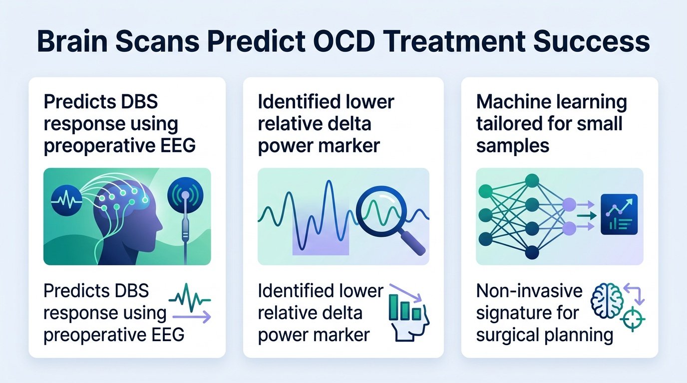
A simple scalp EEG could soon prevent patients from undergoing failed brain surgery for severe OCD.
Deep brain stimulation is a last-resort surgery for severe obsessive-compulsive disorder. It involves drilling holes in the skull to implant electrodes, yet doctors have had no reliable way to know who will actually get better. Patients who do not respond are left with the physical risks of brain surgery and heavy financial debts.
A new study changes this equation. By identifying a specific electrical signature on a standard, non-invasive EEG, researchers can now screen candidates before they enter the operating room. This shifts the clinical approach from a desperate gamble to a calculated, personalized intervention.
The predictive power
The researchers analyzed preoperative resting-state EEG data from a randomized, double-blind, sham-controlled trial of 24 patients. The surgery targeted the nucleus accumbens and the anterior limb of the internal capsule. They discovered that a specific brain wave pattern could reliably predict which patients would benefit from the treatment.
- The marker, characterized by lower relative delta power at a right fronto-temporal electrode, predicted greater symptom reduction at six months.
- This single signature explained more than 40% of the variance in patient outcomes.
- Screening with this method improved the treatment response rate by over 20% compared to standard patient selection.
- The model successfully predicted outcomes in an independent validation cohort of 7 out of 8 patients.
Crucially, the signature only predicted recovery for patients who received active stimulation. Those in the sham stimulation group showed no such pattern. This distinction proves the EEG is measuring a specific capacity to respond to electricity, not just a general tendency to get better over time.
The biological mechanism
This is not a random mathematical correlation. The electrical signature is highly correlated with the right fronto-temporal aperiodic exponent. This links the biomarker directly to the brain’s excitation-inhibition balance, specifically in cortical regions rich in inhibitory-neuron markers.
Furthermore, changes in this signature tracked clinical improvement over time. This means the EEG does not just predict the future. It also serves as a real-time monitor to show if the stimulation is actively engaging the correct brain circuits.
The clinical reality
We must acknowledge the scale of this research. The primary trial cohort involved only 24 patients, and the validation group was limited to 8 individuals. While the machine learning model was specifically designed to handle small sample sizes, these findings must be replicated in larger trials before surgeons can rely on them routinely.
Even with these limitations, the implications are clear. Psychiatry has long chased expensive, complex imaging tools to justify invasive procedures. This study suggests that a cheap, accessible scalp EEG can capture the complex neural architecture required for successful neuromodulation.
Read the full study on medRxiv.