
A new CT-based machine learning model accurately differentiates fresh from old vertebral compression fractures, boosting junior radiologist performance without requiring MRI scans.
Differentiating acute from chronic vertebral compression fractures (VCFs) usually demands magnetic resonance imaging. MRI clearly displays bone marrow edema, but access issues and high costs often slow down care. If computed tomography alone can pinpoint acute fractures, triage becomes much faster.
That shift from MRI dependence to CT radiomics changes operational workflow. Rather than replacing radiologists, this technology acts as a diagnostic buffer that catches subtle fracture patterns on routine scans.
Strong External CT Performance
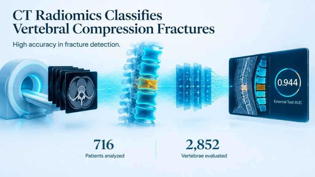
A multi-hospital study analyzed 716 patients (mean age 64.9 ± 9.0 years; 269 males) across 2,852 vertebrae scanned between June 2018 and December 2024. All participants underwent both CT and MRI within one week. Researchers extracted radiomic features from automatically segmented vertebrae to build detection and aging classification models.
The system proved remarkably consistent on unseen data from a separate facility:
- The VCF detection model reached an AUC of 0.944 in the external test set.
- Including an uncertainty category yielded an external classification AUC of 0.892 for distinguishing fresh from old fractures.
- Performance peaked in grade 2 fractures (AUC of 0.891) and crush-type fractures (AUC of 0.911).
These findings align with prior research using machine learning on thoracolumbar CT radiomics to distinguish acute from chronic lesions. Newer approaches also parallel efforts combining deep transfer learning with radiomics for automated spine assessments.
That high baseline accuracy holds across varied fracture types.
Narrowing the Experience Gap
The real clinical value lies in human-AI collaboration. When reading CTs alongside model outputs, clinicians made significantly fewer mistakes. The diagnostic boost proved sharpest for less experienced staff.
AI assistance elevated a junior radiologist’s diagnostic performance from an AUC of 0.628 to 0.793 (p < 0.001). That shift effectively closes the skill gap between junior staff and senior experts during emergency triage.
This decision support prevents unnecessary MRI orders while getting acute fracture patients to treatment faster.
Implementation Reality Check
Retrospective results on closely paired CT and MRI scans do not guarantee identical performance in chaotic emergency rooms. Furthermore, high AUC values on distinct fracture shapes may drop when evaluating mild grade 1 compressions or complex underlying bone disease.
Health systems should view CT radiomics as a clinical multiplier for junior staff rather than an MRI replacement. Prospective trials measuring actual door-to-treatment times remain necessary before cutting MRI protocols entirely.
Read the original research in BMC Medical Imaging.