A new automated pipeline challenges the assumption that high-resolution medical image registration requires massive, slow computing power.
Why do radiologists still spend valuable minutes squinting at side-by-side CT scans to spot kidney tumors? The clinical standard for evaluating renal cell carcinoma relies on subjective visual comparison across different contrast phases. This manual process is slow and prone to human error.
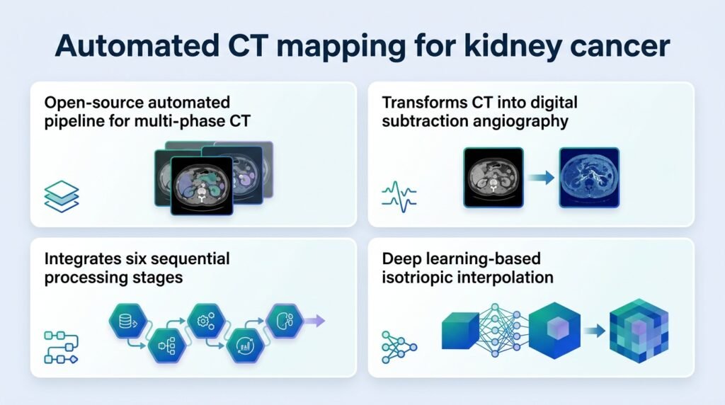
A new pipeline called PRISM (Phase-Resolved Isotropic Subtraction Mapping) aims to automate this by generating clean, color-coded subtraction maps. But the real breakthrough here is not just the automation. It is how the developers solved the computational bottleneck of deformable image registration, proving that smart algorithm design matters more than raw processing power.
The computational shortcut
Deformable registration is notoriously slow. The researchers optimized the pipeline using 200 registrations across five patients. They discovered they could run the heavy calculations on coarse, low-resolution data and apply the results to high-resolution images without losing quality.
Specifically, computing the transform at a coarse 3.0 mm resolution takes only about 4 minutes per phase. The system then applies this transform to high-resolution 0.5 mm volumes. This coarse-register and fine-apply strategy bypasses the typical processing lag entirely.
The team’s systematic parameter testing revealed several highly efficient configurations:
- A 40 mm B-spline grid performed within 6% of the 30 mm quality optimum while cutting computational costs by 36%.
- Using just 5% metric sampling achieved a 3.2x speedup with equivalent quality to 25% sampling.
- A single-level multi-resolution pyramid avoided a 5.4x computational overhead with no quality penalty.
Balancing noise and sensitivity
The pipeline also tackles the delicate balance of image noise. In digital subtraction, setting the right threshold determines whether a doctor sees a tumor or background artifact. The study identified 40 HU (Hounsfield Units) as the optimal subtraction noise threshold.
This 40 HU setting balanced a signal-to-noise ratio of 2.00 against a sensitivity of 14.2% of the enhancing volume retained. Clinicians can adjust this. Higher thresholds of 60-80 HU favor specificity, while a lower 20 HU threshold catches more subtle details.
This precision is crucial for surgical planning. For instance, mapping complex vascular anatomy is vital when evaluating multiple renal arteries before laparoscopic nephrectomy. Better pre-operative imaging directly impacts surgical success.
Why this matters
This shifts the conversation from whether AI can perform this task to whether we can run it in a busy clinic today. PRISM shows that mathematical shortcuts can make advanced 3D vascular mapping practical on standard hardware. This aligns with broader efforts to transition from static 3D imaging to dynamic guidance, similar to the shift toward 4D computed tomography angiography for guiding targeted tumor treatments.
However, the study has a clear limitation. The optimization parameters were tested on just five patients. While the math is sound, clinical validation on a larger, more diverse cohort is required before radiologists can trust these automated subtraction maps for diagnostic decisions.
Read the full study in medRxiv.