
A new graph reasoning model outperforms eye specialists in retinal angiography while showing clinicians exactly how it reached its diagnosis.
Most artificial intelligence tools for eye imaging operate as opaque black boxes. Clinicians routinely hesitate to trust automated software when they cannot trace its logic across the sequential phases of fundus fluorescein angiography (FFA).
That reluctance creates a major barrier to real-world adoption in ophthalmology clinics. Without clear reasoning that matches human clinical logic, even highly accurate models remain sidelined.
A new deep learning framework named FFA-RGNet directly addresses this trust deficit. By explicitly mapping fine-grained lesions across imaging phases and anatomical structures, the model mirrors how human specialists evaluate retinal vascular diseases.
Outperforming Eye Specialists
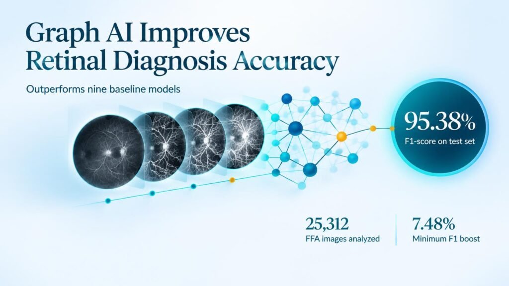
The research team trained and evaluated the model on a multi-center dataset containing 25,312 FFA images from 2,651 patients. On the internal test benchmark, FFA-RGNet achieved a diagnostic F1-score of 95.38% (95% CI: 92.24%–97.88%), comfortably beating nine baseline AI models.
Its diagnostic accuracy surpassed that of qualified ophthalmologists, medical residents, and students across internal, multi-center, and prospective validation sets.
The software delivered its biggest practical value as a clinical decision support tool. When specialists used the framework during image interpretation, their diagnostic F1-scores jumped by 7.48% to 14.11%.
Key Diagnostic Results
- Achieved an F1-score of 95.38% on internal testing.
- Trained on a cohort of 25,312 images from 2,651 patients.
- Boosted human reader F1-scores by 7.48% to 14.11%.
- Outperformed nine baseline AI models in head-to-head evaluations.
Why Traceability Matters
High diagnostic accuracy on static images does not prove a tool is ready to replace human judgment. What sets this framework apart is its structured graph reasoning patterns, which align directly with clinical knowledge to recognize semantic entities across distinct imaging phases.
That clinical alignment provides a clear rationale for every flagged lesion. By showing its work, the software reduces diagnostic uncertainty and helps clinicians catch subtle vascular anomalies without adding overhead to their workflow.
However, retrospective and multi-center validation metrics are not proof of improved patient outcomes. The study evaluated diagnostic accuracy and reader agreement, not whether deployment saves reading time or prevents vision loss in busy clinics.
Until prospective trials demonstrate real-world efficiency gains and better health outcomes, FFA-RGNet serves as a powerful second reader rather than an autonomous diagnostic replacement.
Read the full study in npj Digital Medicine.