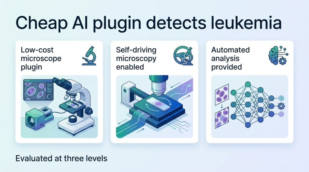
A new hardware plugin bypasses expensive digital scanners to bring automated leukemia screening to basic laboratory microscopes.
For years, digital pathology has faced a major bottleneck. To run advanced diagnostic AI, laboratories must first purchase expensive whole-slide scanners that cost upwards of six figures. This financial barrier effectively locks resource-poor clinics out of modern cancer diagnostics, keeping life-saving technology restricted to wealthy medical centers.
A new system called ALLocate challenges this paradigm. Instead of digitizing entire slides first, this low-cost hardware plugin attaches directly to conventional microscopes to create a “self-driving” system. It analyzes physical glass slides on the fly, skipping the digital scanning step entirely.
How the system works
The engineering shift here is critical. Instead of replacing the microscope, the plugin automates the manual search process. It identifies promising regions, focuses, and classifies cells using onboard AI.
Analyzing bone marrow is notoriously difficult. Unlike simple blood smears, bone marrow contains a highly complex mixture of developing cells. By demonstrating that a basic microscope plugin can navigate this cellular maze with high precision, the researchers have shown that automated triage is possible even for complex hematologic malignancies.
To train the system, researchers used a massive dataset of more than 11,000 annotated regions and 130,000 annotated cells. They then validated the hardware on 165 physical bone marrow smear slides across multiple institutions. This multi-site validation is crucial because bone marrow smears are notoriously difficult to standardize.
The performance breakdown
The diagnostic accuracy holds up remarkably well against traditional, stationary digital scanners. The system achieved high marks across three distinct testing phases:
- An area under the receiver operating characteristic curve (AUC) greater than 0.99 for identifying regions of interest.
- A mean average precision of 0.90 at 50% intersection over union for individual cell detection.
- An overall slide-level diagnostic accuracy of 88% directly on physical glass slides.
That 88% slide-level accuracy is the real story. While it is not a perfect substitute for a board-certified hematopathologist, it provides a highly capable safety net. In regions where specialist expertise is entirely absent, this level of accuracy can reliably flag acute leukemia cases that would otherwise go unnoticed.
The real-world friction
We must be realistic about the limitations. An 88% diagnostic accuracy means some cases will be missed or misclassified without human oversight. This tool is a screening aid, not a final judge. Physical slides also present real-world challenges like dust, poor staining, and preparation artifacts that can confuse the robotic plugin.
Even with these hurdles, the implications are clear. This approach shifts the focus of medical AI from high-end software running on high-end hardware to smart retrofitting. It proves that we do not need to rebuild the lab to upgrade the medicine.
Read the full study in Nature Communications.