AI Classifies Parotid Gland Tumors to Guide Surgery
A new multi-task AI model bridges the diagnostic gap for parotid gland tumors, helping radiologists avoid costly surgical missteps.
Parotid gland tumors present a high-stakes surgical dilemma. If a surgeon misidentifies a benign Warthin tumor as a malignant growth, a patient may undergo an aggressive, unnecessary surgery that risks permanent facial nerve damage. Yet, interpreting ultrasound images of the salivary glands is highly subjective, and misclassifications are remarkably common in daily practice.
This diagnostic bottleneck is where the standard “AI as a second reader” narrative gets complicated. Clinicians do not just need a simple benign-versus-malignant binary. They need precise, multi-class sorting because treatment pathways diverge wildly depending on the specific benign subtype. A new deep learning model called GobletNet addresses this exact complexity by tackling segmentation and four-class classification simultaneously.
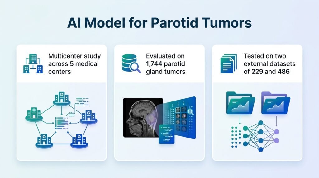
To build a tool capable of handling this nuance, researchers gathered a massive multi-center dataset. The retrospective study analyzed **1,666 patients** with **1,744 parotid gland tumors** across five medical centers. The researchers allocated **1,029 patients** to an internal training set, reserving the remaining cohorts of **229** and **486 patients** for two independent external validation tests.
Surgical stakes and subgroup stability
GobletNet uses a shared encoder to map the boundaries of a tumor while classifying it into one of four categories: malignant tumors, pleomorphic adenomas, Warthin tumors, or other benign lesions. The model maintained high diagnostic accuracy even when tested on external imaging systems it had never encountered before.
- It achieved tumor segmentation Dice similarity coefficients of **0.956** on the internal set, and **0.959** and **0.965** on the two external datasets.
- Classification accuracy reached **0.883** internally, and remained stable at **0.841** and **0.843** in the external validation groups.
- The model achieved one-vs-rest macro-averaged area under the curve (AUC) values of **0.980**, **0.961**, and **0.965** across the three datasets.
- Subgroup analyses proved the model’s performance remains stable across age, gender, and tumor size, with all AUCs remaining **above 0.950**.
The human-AI integration gap
The real value of this technology lies in how it salvages human diagnostic error. In a comparative reader study, GobletNet achieved an overall AUC of **0.962**, easily outperforming every radiologist in the trial. Without assistance, the human readers struggled significantly, logging baseline accuracies between **0.405 and 0.560**.
When radiologists paired their expertise with GobletNet, their diagnostic accuracy jumped to a range of **0.610 to 0.765**. This sharp improvement proves that AI can instantly elevate junior clinicians. However, it also highlights a frustrating bottleneck. Even with a near-perfect algorithm guiding them, human readers still overrode correct machine predictions, keeping the combined accuracy well below the AI’s solo potential.
We must also acknowledge the study’s retrospective design. The algorithm evaluated static, pre-selected historical images rather than real-time, operator-dependent clinical workflows. Prospective clinical trials are still required to prove that these diagnostic gains actually translate to fewer unnecessary surgeries in busy clinics.
Read the full study in BMC Medicine.