 Yusuf Yesil, MD, MIS
Surgeons fighting pancreatic ductal adenocarcinoma often miss extrapancreatic perineural invasion (EPNI) on routine imaging. This microscopic spread along nerve sheaths leads to rapid local recurrence and poorer overall survival. Catching it before surgery changes the operative plan, but human eyes routinely miss the subtle anatomical shifts.
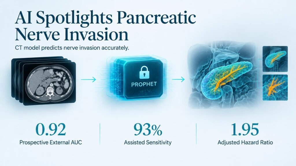
That gap is where an interpretable CT spatial phenotype model steps in. The tool, named PROPHET, calculates risk by examining the spatial relationship between the primary tumor and adjacent abdominal vessels.
High Accuracy Across Multi-Center Cohorts
Researchers evaluated PROPHET across 1,854 patients (median age 63 years; 1,131 male) spanning training, internal testing, neoadjuvant, retrospective external, prospective external, and public dataset validation cohorts. Building on earlier attempts to capture perineural invasion on preoperative CT, this multi-center trial locked its predictive thresholds before running external checks.
The model demonstrated robust diagnostic power across varied clinical scenarios:
- Achieved an AUC of 0.92 in prospective external testing and 0.85 in retrospective external cohorts.
- Maintained an AUC of 0.90 in patients who received neoadjuvant therapy, where tumor margins are notoriously distorted.
- Increased radiologists’ mean sensitivity from 82% to 93% while cutting reading time from 3.64 to 3.02 minutes per patient.
- Stratified overall survival, with high-risk scores linked to shorter survival (adjusted hazard ratio 1.95).
The Specificity Breakdown in Public Datasets
The impressive predictive performance hit a major snag in public benchmark testing. In the Cancer Imaging Archive (TCIA) set, the model yielded an AUC of 0.80, but its specificity collapsed to just 11% (two of 19 cases), despite correctly identifying 80% of positive cases (24 of 30).
That sharp drop highlights a classic AI trap: algorithms tuned on standardized clinical protocols can over-call risk when exposed to heterogeneous image quality. It echoes past concerns over whether post-treatment tissue changes cause CT overestimation of perineural invasion in real-world settings. A tool with high false-positive rates risks over-treating patients or inappropriately denying resection.
While PROPHET significantly accelerates workflow and sharpens radiologist sensitivity, retrospective accuracy is not clinical proof of improved survival. Radiologists should treat high PROPHET scores as a decision support aid, not an independent trigger to alter surgical candidacy.
Read the complete study in Radiology: Artificial Intelligence.