Deploying AI as a routine backstop in radiology departments caught dozens of overlooked liver lesions and forced immediate changes in patient care.
Contrast-enhanced CT scans are the standard tool for spotting liver malignancies, but high-volume workflows create room for human error. A missed lesion on a busy afternoon shift can quietly delay life-saving treatment for months.
That real-world vulnerability is why diagnostic safety nets are moving from theory into practice.
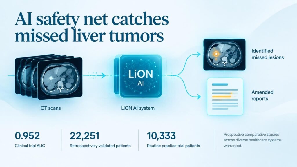
A study published in Nature Medicine evaluates the Liver DiagnOsis Network (LiON), an AI system designed to integrate directly into routine radiology workflows. Rather than trying to replace radiologists, the model sits quietly inside the software ecosystem, analyzing complex multiphase scans alongside clinical data.
High accuracy across complex cohorts
The developers trained LiON on data from 6,443 patients before testing it retrospectively across 22,251 patients in multicenter cohorts. In retrospective testing, the system achieved an area under the receiver operating characteristic curve (AUC) of 0.975.
Diagnostic tools often struggle when underlying organ disease distorts normal anatomy. LiON maintained strong accuracy in complex cases, recording an AUC of 0.971 in patients with hepatic steatosis and 0.924 in those with cirrhosis.
Retrospective accuracy rarely guarantees real-world performance. To test its utility under daily working conditions, researchers ran a single-arm trial following 10,333 patients in routine clinical practice, positioning LiON as an additional automated reader.
Translating software alerts into medical action
The pragmatic trial met its primary endpoint, delivering a real-world AUC of 0.952. More importantly, the trial tracked what happened after the algorithm flagged missed scans.
- The AI-human collaboration identified 51 previously overlooked liver lesions.
- Among those missed findings, 15 were confirmed as true malignancies.
- The secondary findings triggered 37 amended radiology reports.
- The flagged cases led directly to 22 multidisciplinary team escalations and clinical treatment alterations.
That shift in care pathways shows the tangible value of software integration. The algorithm did not just generate diagnostic scores; it actively caught clinical blind spots and prompted doctors to alter treatment plans.
The limit of a single-arm trial
These findings show clear workflow utility, but the study design has key limits. A single-arm trial cannot measure how much extra stress or fatigue false alarms might place on radiologist workflows over time.
It also measures diagnostic accuracy, not patient survival. Catching 15 extra malignancies early is promising, but prospective comparative trials across varied global hospital networks are still required to prove long-term benefit.
For hospital leaders, the takeaway is simple. AI achieves its highest immediate value not by writing the primary report, but by serving as an automated second reader that prevents catastrophic diagnostic drops.
Read the complete study in Nature Medicine.