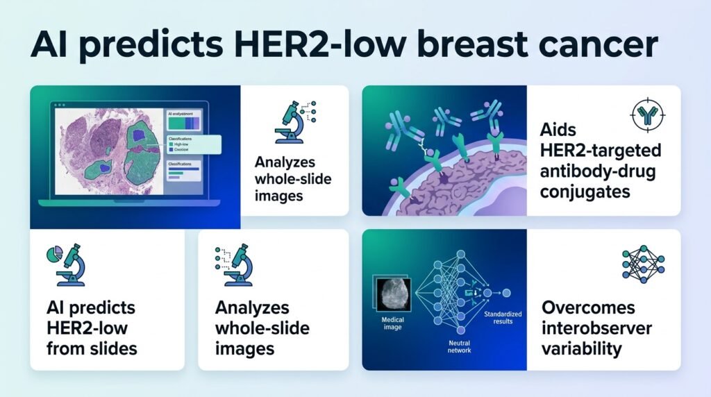
A new deep learning model attempts to spot hard-to-find HER2-low breast cancers from standard tissue slides, but its modest accuracy reveals just how difficult this diagnostic boundary is to draw.
Can an algorithm find what human eyes routinely miss? In breast cancer care, the line between HER2-negative and HER2-low determines who receives life-extending antibody-drug conjugates. Right now, expensive and subjective immunohistochemical tests make that call, often leading to conflicting diagnoses.
This study challenges the assumption that deep learning can easily bypass traditional staining. By using cheap, standard hematoxylin and eosin (H&E) slides, researchers hoped to find a digital shortcut. Instead, the results prove that HER2-low status is too biologically subtle for standard computer vision to master on its own. It forces us to rethink whether visual morphology alone contains enough signal to predict low-level protein expression.
The data behind the model
To build the predictive framework, researchers retrospectively collected 776 cases of invasive breast carcinoma diagnosed at the Affiliated Hospital of Zunyi Medical University between January 2019 and April 2023. They paired an ImageNet-pretrained ResNet50 model for feature extraction with a Clustering-constrained Attention Multiple Instance Learning (CLAM) model. The team used a 10-fold cross-validation method to test the system’s accuracy.
The model’s performance reveals the steep climb ahead for digital pathology:
- The system achieved a mean AUC of 0.613 ± 0.118 on the validation set.
- Accuracy remained low on the test set, yielding a mean AUC of 0.608 ± 0.104.
- Attention heatmaps successfully highlighted predictive regions, giving pathologists visual clues for the model’s choices.
An AUC of 0.608 is only slightly better than a coin flip.
This disappointing performance underscores a harsh reality. HER2-low expression does not leave a distinct structural footprint on standard H&E slides. Earlier attempts, such as a 2020 study in the Journal of Pathology Informatics, also struggled to extract HER2 status from basic tissue stains. While alternative methods like using whole slide gray value maps have attempted to capture these patterns, the biological signal in standard slides remains incredibly faint.
Why this finding matters
This matters because clinicians cannot use this model to make high-stakes treatment decisions. A false negative denies a patient a highly effective therapy, while a false positive exposes them to drug toxicity without benefit. The findings suggest that visual patterns in tumor cells may not correlate directly with low-level protein expression. This disconnect indicates that we may need multi-modal AI models that combine pixel data with genomic sequencing to achieve clinical utility.
The study is limited by its single-center design and small cohort of 776 cases. Until algorithms can reliably cross the 0.80 AUC threshold on external datasets, pathologists must continue to rely on traditional, manual staining.
Read the full study in Diagnostic Pathology.