Low-cost portable MRI scanners can track multiple sclerosis progression, but only if we stop relying on algorithms built for high-end hospital machines.
Can a tiny 64-millitesla magnet on wheels replace a massive 3-Tesla scanner for tracking multiple sclerosis?
The hardware is already here, but the software has been a bottleneck. Standard AI models trained on pristine, high-field hospital scans fail when fed the noisy, low-resolution data from portable machines. This study proves that to make portable neuroimaging viable, we must train AI on the messy reality of ultra-low field data.
The software performance gap
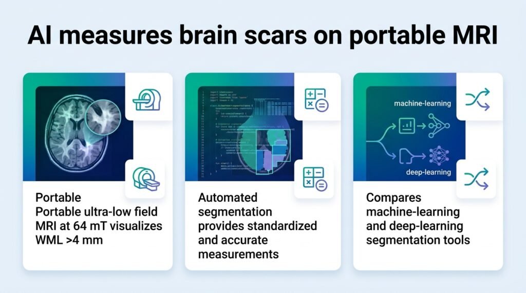
To find the best tool, researchers analyzed same-day paired scans from 84 adults with MS or suspected-MS. The cohort had a mean age of 48 years and included 62 females. They tested six automated segmentation methods against manual annotations on portable ultra-low field (pULF) scans. The results exposed a massive performance gap between generic models and those tailored for low-field hardware.
- The PLAn-FL model, pre-trained on high-field scans and refined on 64mT data, achieved the highest Dice score of 0.50 ± 0.24.
- Generic models like MIMoSA struggled, scoring just 0.24 ± 0.20.
- The widely-used WMH-SynthSeg scored 0.30 ± 0.18, while standard nnU-Net-FL reached 0.41 ± 0.24.
- Except for MIMoSA, all advanced models generated lesion volumes that correlated significantly with patient disability scores.
PLAn-FL succeeded because it used a two-step training process. It first learned what lesions look like from pristine high-field scans, then adapted to the noisy 64mT environment. This hybrid approach is the blueprint for future clinical AI.
Redefining clinical trial access
This is not about replacing high-field MRIs for initial diagnosis. It is about decentralizing clinical trials and routine monitoring. If a portable machine in a rural clinic can track brain lesions accurately enough to correlate with physical disability, we can run larger, more diverse clinical trials.
These disability metrics, the Expanded Disability Status Scale (EDSS) and Scripps Neurologic Rating Scale (SNRS), are the gold standards for tracking MS. The fact that the AI-derived volumes from PLAn-FL correlated with these scores after adjusting for age is the real clinical win. It means the software is tracking real-world patient decline, not just digital noise.
This builds on previous work showing that blinded readers can detect MS lesions on portable scanners, such as the findings in the High‐Field‐Blinded Assessment of Portable Ultra‐Low‐Field Brain MRI.
However, we must be honest about the limitations. A Dice score of 0.50 means the AI still misses or misidentifies half of the lesion volume compared to human experts. Portable scanners only reliably visualize lesions larger than 4 mm. For tracking micro-active disease, high-field scanners remain indispensable.
Read the full preprint on medRxiv.