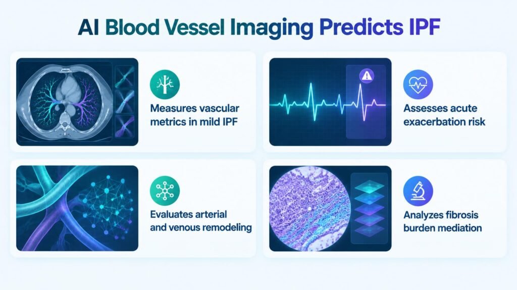
A new deep-learning analysis reveals that blood vessel changes, not just scarring, drive sudden and deadly lung failures in patients with mild disease.
Clinicians treating mild idiopathic pulmonary fibrosis (IPF) often rely on a false sense of security. Patients with stable lung capacity can suddenly suffer fatal lung failures, leaving doctors with few warning signs. This analysis challenges the traditional clinical focus on lung scarring alone by pointing to the critical, hidden role of blood vessels.
The clinical consequence is immediate. By analyzing blood vessels instead of just scars, clinicians can identify high-risk patients before their lung function drops. This shifts the focus from late-stage damage control to proactive monitoring. It forces a rethink of how we stage mild lung disease.
The hidden vascular driver
The retrospective study tracked 313 patients with mild IPF, defined by a forced vital capacity of 80% or higher and a diffusing capacity for carbon monoxide of 55% or higher. This cohort had a mean age of 66.4 years and included 245 male patients. Over a median 6.3 years of follow-up, 86 patients (27.5%) suffered an acute exacerbation, 2 patients (0.6%) underwent lung transplantation, and 120 patients (38.3%) died.
The deep-learning software proved that the damage from lung scarring is heavily mediated by vascular remodeling. Specifically, venous remodeling mediated 57.1% of the fibrosis effect on acute exacerbations. Meanwhile, small-artery volume fraction mediated 21.5% of this effect. Looking only at scars misses the active vascular remodeling that actually triggers sudden patient decline.
Predicting sudden patient decline
The software extracted precise vascular metrics from baseline unenhanced chest CTs to predict patient outcomes. The key findings include:
- Venous dilatation increased the risk of acute exacerbation by an adjusted hazard ratio of 7.93.
- Venous diameter heterogeneity raised the risk of exacerbation by an adjusted hazard ratio of 1.37.
- A higher small-artery volume fraction was protective, reducing the risk of exacerbation by an adjusted hazard ratio of 0.91.
- A higher fibrosis score was associated with a higher risk of acute exacerbation, with an adjusted hazard ratio range of 1.05 to 1.09.
- A higher fibrosis score was also associated with a higher risk of the composite outcome of exacerbation, transplant, or death, with an adjusted hazard ratio range of 1.08 to 1.20.
Moving beyond single-site data
These results come from a retrospective study at a single tertiary hospital. Because of this, we cannot assume these AI metrics will perform the same way across different scanner types or diverse patient populations. It is a predictive association, not a proven tool to replace clinical judgment yet.
However, the practical takeaway is clear. Radiologists should look beyond the sheer volume of lung scarring. By integrating automated vascular measurements into routine CT staging, clinics can flag high-risk patients with supposedly mild disease for early, aggressive monitoring.
Read the full study in Radiology.