The field of pulmonology is undergoing a structural transition as artificial intelligence moves beyond simple computer-aided detection toward multimodal prognostic systems. Historically, clinical AI in respiratory medicine was confined to binary classification tasks, such as identifying a nodule on a chest radiograph or segmenting a lung lobe on a computed tomography (CT) scan. Today, the integration of clinical variables, histopathology, genomics, and longitudinal imaging is enabling clinicians to predict disease progression, therapeutic response, and structural decline with unprecedented precision. This shift is particularly critical in oncology and interstitial lung diseases, where early intervention directly correlates with survival outcomes.
This month’s developments highlight a clear trend: regulatory bodies and clinical researchers are validating tools that extract subclinical signals from routine diagnostic tests. Whether identifying pulmonary hypertension risk from a standard 12-lead ECG or utilizing frozen vision foundation models to characterize early-stage lung adenocarcinoma, AI is proving its utility as a clinical force multiplier. For clinicians, digital-health investors, and product teams, the challenge is no longer demonstrating that AI can see abnormalities, but proving that AI-driven insights translate into measurable improvements in patient management and clinical trial efficiency.
Notable papers
• Imaging-based development and validation of artificial intelligence models for lung adenocarcinoma precursor lesions and early lung adenocarcinoma presenting as pulmonary nodules
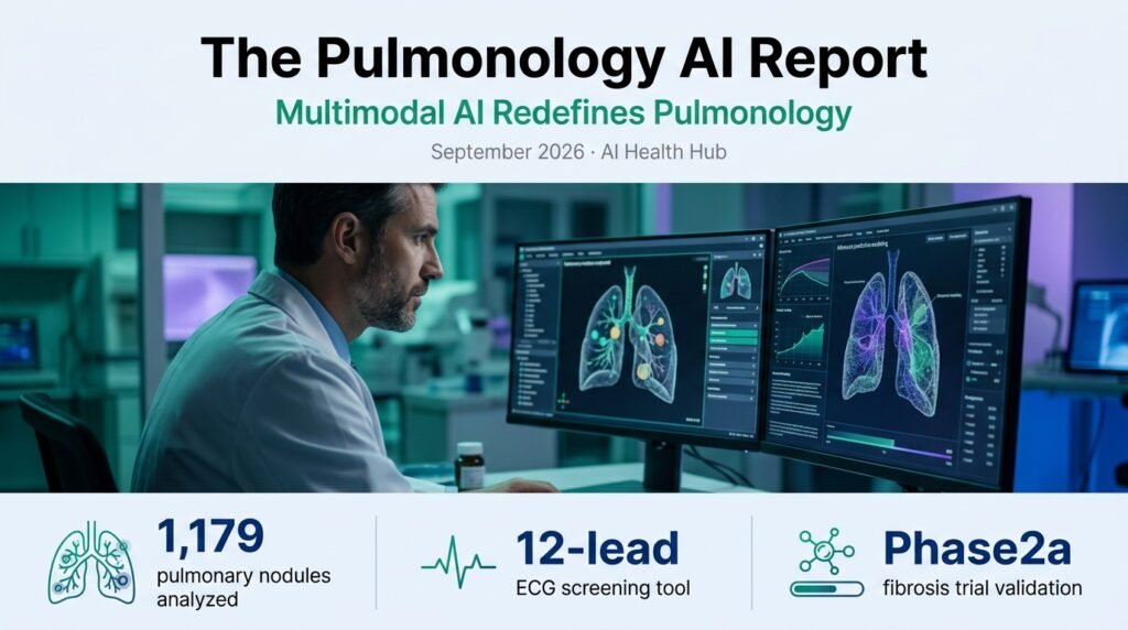
This retrospective single-center study developed and internally validated a multimodal framework integrating CT representations from a frozen vision foundation model with clinical variables, evaluating 1,179 pathologically confirmed pulmonary nodules to distinguish precursor lesions from invasive early lung adenocarcinoma.
Brand take: Genuinely useful clinically because distinguishing pre-invasive lesions from invasive adenocarcinoma on CT scans prevents unnecessary surgical resections while ensuring early intervention for aggressive malignancies.
• A multistage CT-based artificial intelligence system for microvessel density quantification in lung adenocarcinoma: correlation with histopathology
Researchers developed and validated a multistage CT-based AI system to noninvasively quantify microvessel density (MVD) in 152 surgically resected lung adenocarcinoma patients, demonstrating a strong correlation with CD34-immunostained histopathological microvessel counts.
Brand take: Underrated because noninvasive quantification of tumor angiogenesis via routine CT imaging could serve as a powerful biomarker for anti-angiogenic therapy selection without requiring repeat biopsies.
• Prognostic value of AI-derived probability of interstitial lung abnormalities and interstitial lung disease in patients with esophageal cancer
This retrospective study evaluated the association between AI-derived probability of interstitial lung abnormalities (ILA) and interstitial lung disease (ILD) in an esophageal cancer cohort re-evaluated according to the 2025 American Thoracic Society (ATS) Clinical Statement, finding that AI-derived ILA probability is a strong independent predictor of poor overall survival.
Brand take: Genuinely useful clinically as it allows oncologists to identify subclinical pulmonary fibrosis before initiating cardiotoxic or pneumonitis-inducing cancer therapies.
• A Computational Histology Artificial Intelligence Prognostic Biomarker in Non-Small Cell Lung Cancer Using the Cancer Genome Atlas
This study evaluated whether a Computational Histology Artificial Intelligence (CHAI) biomarker, derived solely from diagnostic hematoxylin-and-eosin (H&E) whole-slide images of non-small cell lung cancer (NSCLC) patients, could provide prognostic information independent of established clinicopathologic factors.
Brand take: Overhyped because clinical translation of H&E-only prognostic models remains heavily bottlenecked by pre-analytical staining variations across different pathology laboratories.
• Patient preferences for the use of AI in chest X-ray result processing and communication
Using the Technology Acceptance Model and Health Belief Model, this study evaluated patient preferences regarding the use of AI in chest X-ray processing and communication, finding that while patients accept AI as a supportive tool, they strongly reject automated communication of abnormal results without direct physician oversight.
Brand take: Underrated because patient trust and communication preferences are the ultimate gatekeepers of real-world clinical AI adoption and workflow integration.
• Caregivers’ experiences of using an artificial intelligence-based system for paediatric asthma family management: a qualitative study
This qualitative study investigated how an AI-based system designed for pediatric asthma family management influenced everyday caregiving, highlighting that while the system improved disease knowledge, caregivers experienced technical friction and alert fatigue.
Brand take: Overhyped because consumer-facing digital health tools frequently fail to maintain long-term caregiver engagement due to a lack of integration with primary care workflows.
Products, deals & funding
• Brainomix & Endeavor Clinical Data
Brainomix and Endeavor BioMedicines presented clinical data at the ERS Congress 2026 demonstrating that the AI-powered ‘e-Lung’ CT analysis tool successfully detected significant improvements in lung structure and fibrosis in a Phase 2a trial of taladegib for patients with idiopathic pulmonary fibrosis (IPF).
Brand take: Genuinely useful clinically as it validates AI-derived imaging endpoints as sensitive, objective markers of therapeutic efficacy in respiratory clinical trials.
• Luca Healthcare ERS Presentations
Luca Healthcare presented two validation studies at the ERS Congress 2026, demonstrating the clinical utility of its AI-powered cough analysis for community-based COPD screening and its ‘DeepSpiro’ smartphone-based lung function estimation technology.
Brand take: Underrated because smartphone-based spirometry and acoustic analysis democratize access to pulmonary screening in low-resource settings where traditional spirometry is unavailable.
• Thirona Strategic Shift
Thirona announced a major strategic divestment, selling its retinal AI division to Revenio Group to fully focus its engineering and commercial resources on AI-driven lung imaging for pulmonary precision medicine and interventional pulmonology.
Brand take: Genuinely useful clinically because specialized focus will accelerate the development of high-fidelity 3D bronchoscopy guidance and personalized therapeutic planning tools.
Regulatory & clinical adoption
• DeepHealth FDA Clearance
DeepHealth received FDA 510(k) clearance for its foundation model-based Chest X-Ray AI solution, which detects and localizes major thoracic abnormalities, including nodules, pleural effusion, and consolidation.
Brand take: Genuinely useful clinically as foundation models improve generalization across diverse hospital imaging systems, reducing false-positive rates in high-throughput settings.
• Tempus ECG-PH FDA Clearance
Tempus AI received FDA 510(k) clearance for Tempus ECG-PH, an AI-enabled software device that analyzes standard 12-lead ECGs to identify patients at high risk for pulmonary hypertension, a condition notoriously difficult to diagnose early.
Brand take: Underrated because screening for pulmonary hypertension via routine ECGs can drastically shorten the multi-year diagnostic odyssey these patients typically face.
• Oncology Society of Chinese Medical Association Guideline
The Oncology Society of the Chinese Medical Association released its 2026 clinical diagnosis and treatment guideline for lung cancer (https://doi.org/10.3760/cma.j.cn112137-20260511-01255), standardizing early detection and treatment pathways and formally incorporating advanced imaging and AI-assisted screening protocols.
Brand take: Genuinely useful clinically as national guidelines begin to formalize the role of AI in early-stage lung cancer detection and management.
Trends & what to watch
The pulmonology AI landscape is rapidly shifting from diagnostic assistance to therapeutic guidance. The validation of Brainomix’s ‘e-Lung’ platform in a Phase 2a trial indicates that pharmaceutical developers are increasingly relying on AI-driven quantitative CT analysis to measure treatment response in interstitial lung diseases. This trend is expected to accelerate over the next 1-3 months, with more drug developers integrating AI imaging biomarkers into their clinical trial protocols to detect micro-structural changes that traditional forced vital capacity (FVC) measurements might miss.
Concurrently, the regulatory landscape is favoring opportunistic screening tools. The FDA clearance of Tempus ECG-PH demonstrates a growing interest in using AI to extract secondary diagnostic value from common, low-cost tests. By analyzing standard 12-lead ECGs for signs of pulmonary hypertension, clinical systems can flag at-risk patients during routine cardiovascular workups. Product teams should focus on developing similar opportunistic screening algorithms that leverage existing, underutilized clinical data to identify silent pulmonary pathologies before they progress to advanced stages.
Bottom line
Pulmonology AI has matured from basic diagnostic image readers into multimodal prognostic engines capable of predicting therapeutic response and identifying subclinical disease from routine clinical tests.