
Standardizing Fetal Ventriculomegaly Diagnosis
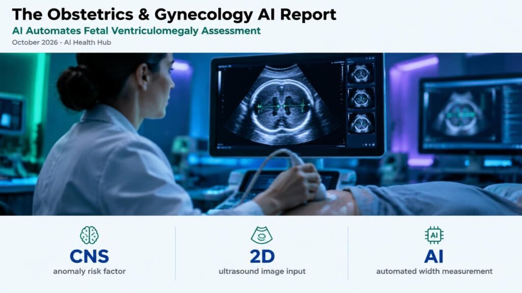
Fetal ventriculomegaly is one of the most common central nervous system abnormalities detected on prenatal ultrasound, serving as a critical indicator for underlying congenital defects, chromosomal anomalies, and intrauterine infections. Accurate measurement of the fetal lateral atrium width is required to stratify risk and guide prenatal counseling. However, manual ultrasound assessment remains highly operator-dependent, leading to measurement variability between clinicians and potential misclassification of borderline cases.
To address these diagnostic inconsistencies, researchers evaluated a deep learning framework designed to automate the measurement and diagnosis of fetal ventriculomegaly from standard ultrasound images (BMC Medical Imaging). The model automatically segments the lateral ventricles, identifies correct anatomical planes, and measures ventricular width, establishing an objective diagnostic threshold without requiring manual caliper placement by the sonographer.
Diagnostic Consistency and Clinical Workflow Impact
By automating plane verification and measurement, the deep learning tool aims to reduce intra- and inter-observer variability across clinical settings. In routine obstetric practice, minor differences in manual caliper placement can shift a diagnosis between normal variation and mild ventriculomegaly, generating patient anxiety or triggering unnecessary downstream invasive testing. Automated measurement provides standardized outputs that support less experienced sonographers and streamline expert review in tertiary care centers.
Key Limitations and Practical Guidance
Despite these automated precision gains, important clinical limitations temper immediate full autonomy. The model relies heavily on image quality and clear visualization of anatomical landmarks; acoustic shadowing, maternal body habitus, or suboptimal fetal positioning can impair segmentation accuracy. Furthermore, automated lateral ventricle measurement functions strictly as a diagnostic support tool rather than a standalone clinical decision maker. Clinicians must still correlate automated measurements with comprehensive anatomical surveys, detailed neurosonography, and maternal serum or genomic evaluations. For maternal-fetal medicine specialists and sonographers, integrating deep learning algorithms into ultrasound systems should focus on preliminary screening and quality assurance, ensuring human expert oversight remains central to final diagnostic reporting.