A new machine learning model proves that routine CT scans hold hidden data capable of predicting brain hemorrhage outcomes far better than human clinical judgment alone.
Clinicians treating spontaneous intracerebral hemorrhage (ICH) have long relied on basic physical measurements like hematoma volume to guess a patient’s trajectory. This approach is notoriously imprecise. It leaves families and care teams in limbo during critical early hours when decisive action is needed most.
By extracting quantitative features from standard non-contrast CT (NCCT) scans, researchers have demonstrated that mathematical texture analysis can turn standard imaging into a highly accurate prognostic engine. This challenges the status quo of relying on visual inspection. It suggests that clinical decisions should be guided by quantitative imaging signatures that the human eye cannot detect. This shift directly impacts clinical triaging and family counseling by replacing subjective clinical intuition with objective, data-driven forecasting.
This builds on previous research showing that radiomics scores can predict hematoma enlargement. However, this new model takes the science a step further by directly forecasting overall functional recovery and patient risk stratification.
The predictive leap
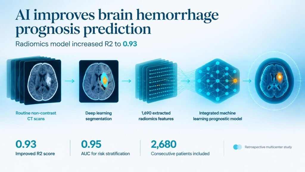
The retrospective study analyzed a large multicenter cohort of 2,680 consecutive patients. The researchers trained their system on 1,876 cases, ran internal validation on 804, and tested it externally on 196 patients. The deep learning model automatically segmented the hematomas and extracted 1,690 radiomics features from admission scans, eventually isolating 42 highly reproducible features to build a consolidated “Rad-score.”
When combined with basic clinical data, the radiomics-driven model vastly outperformed traditional clinical-only assessments. The performance gap between the old clinical methods and the new integrated model is stark:
- For functional outcome prediction, the integrated model boosted the R2 metric from 0.35 to 0.93, while slashing the mean squared error from 387 to 61.
- For prognostic risk stratification, the clinical-only model managed a poor AUC of 0.59, whereas the fully integrated model achieved 0.95.
- In independent external validation, the integrated model maintained a robust AUC of 0.86, proving it can generalize beyond its original hospital setting.
The path to adoption
While these numbers are impressive, the study relies on retrospective data. Retrospective performance does not guarantee real-world clinical utility, and the model has not yet been shown to improve actual patient survival rates in a live trial. Furthermore, the researchers noted an intriguing, unproven link between ambient temperature at symptom onset and disease progression. This variable requires prospective validation before it can be trusted.
For healthcare leaders, the takeaway is clear. Do not wait for entirely new imaging hardware to improve stroke care. The immediate opportunity lies in deploying software that extracts deeper insights from the basic NCCT scanners already sitting in every emergency department. This aligns with earlier validation efforts showing that radiomic markers outperform visual markers for predicting patient decline. Hospitals should focus on integrating these automated quantitative tools into existing radiology workflows to assist, rather than replace, clinical decision-making.
Read the full study in medRxiv.