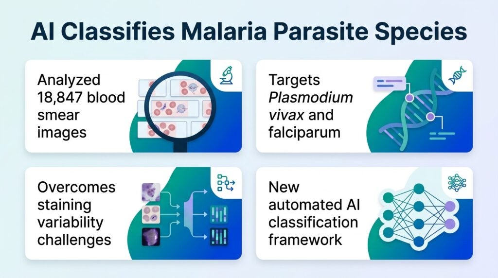
A new hierarchical deep learning framework separates malaria species from healthy cells with near-perfect accuracy, even when clinical data is highly imbalanced.
Can a machine tell the difference between two microscopic killers that look almost identical under a lens? For clinicians treating malaria, mistaking Plasmodium vivax for Plasmodium falciparum can lead to incorrect drug prescriptions and fatal outcomes. Yet manual microscopy remains slow, subjective, and highly dependent on the skill of the technician.
This diagnostic bottleneck is where automation usually stumbles.
A new study challenges the assumption that diagnostic AI requires perfectly balanced, pristine datasets to be clinically useful. Most deep learning models fail when they encounter rare cases in the wild. By using a hierarchical approach—first spotting infected cells, then classifying the species—this model proves that smart architecture can overcome the messy reality of clinical data.
How the model works
Researchers trained the system on 18,847 Giemsa-stained thin blood smear images from the Indian Council of Medical Research–National Institute of Malaria Research. The dataset included uninfected red blood cells alongside cells infected with either P. vivax or P. falciparum. To find the most robust setup, the team evaluated the framework using three different train-validation-test splits: 60:20:20, 70:15:15, and 80:10:10.
This hierarchical strategy paid off. Instead of trying to solve the entire classification problem at once, the AI first separated healthy cells from sick ones. Only then did it attempt the harder task of distinguishing the two parasite species.
The performance breakdown
The model delivered high marks across the board, showing remarkable resilience when data was scarce.
- Using the 70:15:15 split, the framework achieved 98.20% accuracy and a 97.54% F1-score.
- The Area Under the Receiver Operating Characteristic (ROC-AUC) reached 99.66%.
- The Matthews correlation coefficient, which penalizes lucky guesses on imbalanced data, hit 95.42%.
- At a highly imbalanced 2% training prevalence, the model still maintained 80.61% accuracy and a 76.61% F1-score.
The real-world catch
This resilience is the real story here. In actual clinics, infected cells are rare compared to healthy ones. A model that only works in a perfectly curated lab is useless in the field. This framework’s ability to maintain utility at 2% prevalence means it can actually handle the skewed data of real-world triage.
However, we must be honest about the limitations. The model was trained on high-quality images from a centralized national institute. Real-world clinics often use poorly maintained microscopes and inconsistent staining techniques. If the input quality drops, that high accuracy will likely degrade. Furthermore, the study did not test the model against other malaria species like P. ovale or P. malariae, which limits its global utility.
Even with these caveats, this framework represents a practical step forward. It shows that diagnostic tools do not need perfect data to deliver reliable answers.
Read the full study in BMC Medical Informatics and Decision Making.