
A foundation model reading routine heart traces can flag dementia risk up to a decade before diagnosis, but it is a triage filter, not a standalone diagnostic tool.
Age remains the single largest risk factor for dementia. That reality makes it difficult for predictive algorithms to prove they are finding genuine biological signals rather than simply guessing how old a patient is.
A new study published in the Journal of Medical Internet Research tests whether AI foundation models reading routine electrocardiograms can break through that barrier. The findings confirm that heart traces carry age-independent signals of cognitive decline, though the strength of that signal demands realistic expectations about how clinicians should use it.
Testing the Heart-Brain Connection
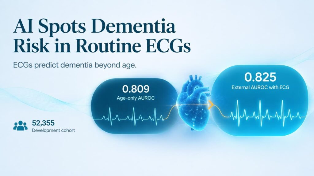
Researchers evaluated a knowledge-enhanced foundation model using retrospective data from two Korean hospitals. The primary development cohort at Severance Hospital included 52,355 patients, while external validation was conducted on 6,500 patients at Yongin Severance Hospital. The analysis specifically focused on routine 12-lead ECGs interpreted as normal sinus rhythm to predict cognitive impairment or dementia within the following 10 years.
Chronological age alone established a high baseline for risk prediction, reaching an external AUROC of 0.809. Fusing age with the model’s ECG-derived probability pushed the external AUROC to 0.825, producing statistically significant improvements in calibration and net reclassification benefit.
To prove the software was not merely re-learning patient age, the team stripped out the age-predictable component. The remaining age-residualized embedding maintained a modest but reproducible signal, yielding an internal AUROC of 0.590 and an external AUROC of 0.579.
Tracking the Prediagnostic Window
The electrophysiological baseline did not stay static over time. By tracking phenotypic features across four distinct prediagnostic time frames, researchers watched subtle cardiac shifts emerge long before clinical cognitive testing occurred.
- 52,355 development and 6,500 external validation patients were analyzed across two hospital sites.
- Adding ECG probabilities to patient age increased external predictive accuracy from 0.809 to 0.825 AUROC.
- Age-independent heart embeddings retained a standalone predictive signal of 0.579 AUROC.
- Divergence in cardiac patterns appeared primarily within 1 to 3 years of formal diagnosis.
Out of 105 evaluated ECG labels, 19 (18.1%) showed significant divergence between cases and matched controls. These changes were dominated by nonspecific ST-segment and T-wave alterations, acute ischemic patterns, and atrial arrhythmias. Most of this separation concentrated in the 1 to 3 years and 0 to 1 year windows before formal diagnosis.
Triage Filter, Not Diagnostic Tool
These findings challenge the idea that early dementia detection requires expensive scans or invasive biomarkers right out of the gate. Routine heart traces taken during standard physicals or minor surgeries already capture parallel vascular and neural aging.
However, the data clearly argues against relying on software to replace cognitive evaluations. An age-residualized AUROC of 0.579 is far too weak to justify a primary diagnosis. Furthermore, because the study relied on retrospective East Asian cohorts and excluded abnormal rhythms, real-world prospective performance across diverse care settings remains unproven.
The actionable value lies in low-cost screening. Combining routine ECG readings with basic age profiles gives primary care networks an automated alert system to flag patients who need formal neurological workups before overt cognitive failure occurs.
Read the full study in the Journal of Medical Internet Research.