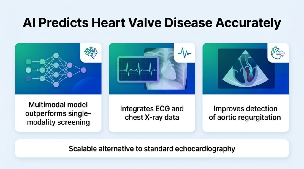
By fusing electrical and structural data, a new multimodal model flags hidden heart valve risks before symptoms appear.
Why do we wait for a patient’s heart valve to fail before we scan it? Echocardiograms are the gold standard for diagnosing leaky valves, but they are too resource-intensive for routine screening. This leaves thousands of patients walking around with silent, progressive heart damage.
The standard clinical playbook relies on single-test shortcuts. Doctors use either an electrocardiogram (ECG) or a chest X-ray to guess at structural heart issues. This study challenges that siloed approach. By forcing these two cheap, ubiquitous tests to talk to each other through AI, researchers bypassed the need for early, expensive imaging.
A massive clinical dataset
The retrospective multicenter study analyzed 212,888 paired ECG and chest X-ray examinations. These came from 116,380 patients across two Chinese clinical centers. Every patient had both tests performed within 60 days of an echocardiogram. The researchers built a neural network to predict progression to moderate-to-severe aortic regurgitation (AR), mitral regurgitation (MR), or tricuspid regurgitation (TR).
The results show a clear advantage for the combined approach:
- For aortic regurgitation, the C-index rose from 0.616 with ECG alone to 0.713 using the multimodal model, achieving an AUROC of 0.729 and an AUPRC of 0.972.
- Mitral regurgitation prediction reached a C-index of 0.801 and an AUROC of 0.814, beating both ECG-only at 0.782 and X-ray-only at 0.775.
- Tricuspid regurgitation prediction achieved a C-index of 0.802, where the combined model offered a higher net clinical benefit on decision curve analysis than single-modality tools.
Why this finding matters
This is not just about slightly better math. It proves that electrical signals and physical shadows contain overlapping, complementary data that humans cannot manually synthesize. If we can catch aortic regurgitation early, we can intervene before the heart muscle permanently stretches and fails.
The AI did not just guess. Interpretability maps showed the model focused on biologically logical areas. It targeted specific ECG leads like II and the precordial leads, alongside physical signs of pulmonary congestion and chamber enlargement on the X-rays. This transparency is crucial for clinical buy-in.
The hurdles ahead
We must be honest about the limitations. The data comes entirely from two centers in China, so we do not know if these algorithms will perform as well on different patient populations. Furthermore, the model requires both tests to be taken within a tight 60-day window, which may not match real-world clinical workflows where tests are ordered sporadically.
Even with these hurdles, the study shows that we do not need exotic new hardware to improve cardiac screening. We just need to connect the cheap tools we already have.
Read the full study in medRxiv.