Title: AI identifies lung cancer subtypes before surgery
A new multimodal AI model can classify lung adenocarcinoma subtypes from CT scans before a surgeon ever makes an incision.
How do you treat a lung tumor when you do not know its exact subtype? Surgeons often have to operate first and ask questions later. Biopsies can miss the most aggressive parts of a tumor, leading to under-treatment or unnecessary lung tissue loss.
This study complicates the traditional reliance on post-operative pathology. By fusing clinical data, peritumoral features, and deep learning, the researchers show we can predict the exact subtype of lung adenocarcinoma before surgery. This shifts the clinical paradigm from reactive pathology to proactive, personalized surgical planning.
Previous efforts often struggled to distinguish subtle histological differences using standard imaging. For instance, earlier models successfully classified general lung cancer histology as seen in Deep learning classification of lung cancer histology using CT images, but pinpointing specific adenocarcinoma subtypes remained a hurdle. Another study compared capsule networks and radiomics to identify subtypes in Identifying the histologic subtypes of non-small cell lung cancer with computed tomography imaging. This new model builds on those foundations by looking beyond the tumor boundary.
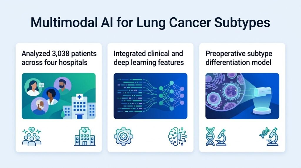
The researchers built and validated their model using a massive multi-center dataset. They gathered data from 3,038 patients across four hospitals. They split this cohort into a training set of 1,822 patients, a test set of 608 patients, and an independent validation set of 608 patients. Two radiologists manually segmented the two-dimensional tumor regions on CT scans. The team then extracted radiomic and deep learning scores, combining them with clinical features selected via the Boruta algorithm and recursive feature elimination.
How the model performed
- The fused model achieved an Obuchowski index of 0.85 in the training set, 0.81 in the test set, and 0.79 in the validation set.
- For the lepidic subtype, F1-scores were 0.77 (training), 0.72 (test), and 0.74 (validation).
- For the acinar/papillary subtype, F1-scores were 0.61 (training), 0.57 (test), and 0.54 (validation).
- For the solid/micropapillary subtype, F1-scores reached 0.63 (training), 0.59 (test), and 0.52 (validation).
The explainability factor
AI in oncology is often criticized as a black box. Doctors cannot risk patient lives on an unexplainable algorithm. To solve this, the researchers used Shapley additive explanations (SHAP) and individual conditional expectation (ICE) plots.
The SHAP analysis revealed that a ResNet-101 feature was the strongest predictor of subtype. This was closely followed by the peritumoral radiomic score and tumor lobulation. This means the tissue immediately surrounding the tumor holds critical clues that human eyes routinely miss.
However, the model is not perfect. The drop in F1-scores for the solid/micropapillary subtype down to 0.52 in the validation set shows that aggressive subtypes remain difficult to classify. Manual segmentation of CT scans by two radiologists is also time-consuming and limits real-world scalability. Automated segmentation must be perfected before this enters routine clinical workflows.
Even with these hurdles, the study proves that preoperative radiomics is moving from academic curiosity to clinical utility. It gives oncologists a non-invasive roadmap to tailor surgeries, potentially sparing patients from over-treatment.
Read the full study in Quantitative Imaging in Medicine and Surgery.