
Radiologists reading low-dose CT scans routinely miss small airway nodules, delaying critical lung cancer interventions.
A unassisted radiologist catches fewer than half of these central lesions. When an algorithm acts as a second reader, that dynamic flips immediately. The clinical hurdle has long been central lesion characterization, where software struggles alongside humans. This multi-center study shows that deep learning can double human sensitivity without clogging the workflow.
That shift matters because early central airway detection directly influences surgical candidacy. Catching a central mass before complete bronchial obstruction alters the entire treatment timeline.
AI boosts radiologist detection rates
Researchers built a deep learning system by adapting a sphere-based center-points matching network with multiscale attention. The team trained the detection pipeline on 907 proximal airway nodules and validated it on an independent multi-center testing set of 738 proximal nodules across four hospitals.
For lesion characterization, a 3D SEResNeXt101 architecture was trained, validated, and tested on 286 airway nodules (121 benign, 165 malignant) using a standard 6:2:2 split before evaluation on an independent test cohort.
The resulting performance numbers showcase clear human-AI synergy:
- Standalone AI detection reached 73.7% sensitivity on the independent multi-center test set.
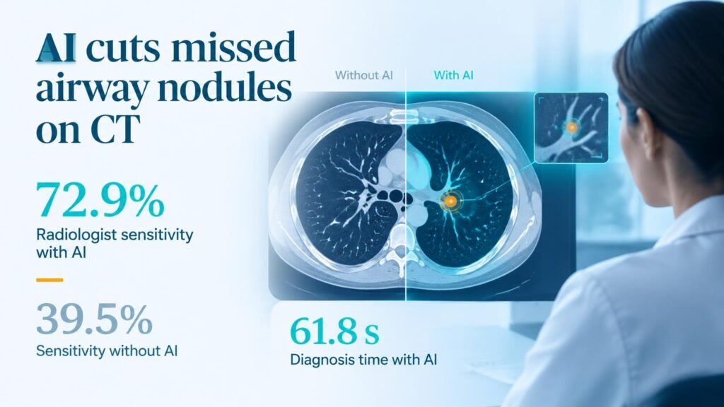
- AI assistance raised radiologist sensitivity from 39.5% to 72.9%.
- Human-AI reader pairing boosted the F1-score from 49.4% to 77.2%.
- Mean diagnostic time per case dropped from 83.8 seconds to 61.8 seconds.
- Malignancy classification sensitivity reached 72.7% with a 87.5% specificity on the independent test set.
Workflow gains meet operational realities
These findings build on broader efforts to automate thoracic imaging, such as combining YOLO and CNN architectures for lung tumor segmentation. They also complement recent work mapping how AI guides downstream diagnostic bronchoscopy.
The drop in reading time by 22 seconds per scan disproves the common objection that secondary AI readers slow clinicians down. Software that adds review steps usually creates friction. Here, clear target highlights streamlined reader focus.
This operational gain proves that concurrent software can shave seconds off routine reads while catching critical missed pathology.
The catch behind the numbers
Retrospective multi-center evaluations still fall short of proving downstream patient survival gains. The classification model showed modest specificity in initial validation sets, dipping to 62.7% in early training splits before stabilizing.
A elevated false-positive rate forces follow-up procedures that carry cost and anxiety. Segmental airway anatomy also presents motion artifacts that clean retrospective datasets rarely reflect.
Health systems should treat this tool strictly as a concurrent second reader rather than an automated triage filter. The real test comes when deployed in high-volume screening programs, where reader fatigue is real and real-world image quality varies widely.
Read the full paper in Insights into Imaging.