
Relying on artificial intelligence to clear stroke patients can leave deadly arterial blockages completely undetected.
A negative scan result from stroke AI does not mean the brain is safe. While algorithm vendors frequently advertise high overall accuracy, anatomical location dictates clinical utility. When software evaluates the main arterial trunks, it functions reliably. But deep in the vascular tree, its diagnostic value collapses.
That blind spot poses an immediate risk in emergency triage. Clinicians who treat a negative AI alert as a green light to rule out intervention risk missing treatable distal clots. This meta-analysis forces a hard boundary on how hospitals deploy triage software: AI works as an emergency bell for primary blockages, not a diagnostic safety net for subtle ones.
A Tale of Two Vessels
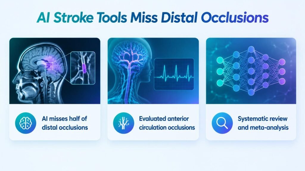
A systematic review in Radiology: Artificial Intelligence analyzed 31 reports encompassing 15,708 patients to evaluate CT angiography (CTA)-based stroke algorithms across distinct anatomical zones. The aggregate numbers mask a dramatic drop in sensitivity depending on vessel depth.
Early benchmarks in stroke AI validation established strong overall performance. However, breaking performance down by anatomy paints a vastly different picture:
- Global LVOs: Pooled sensitivity reached 80.1% with a specificity of 91.9%.
- Proximal Occlusions (ICA/M1): AI caught major blockages effectively, achieving 91.6% sensitivity and 92.7% specificity with moderate evidence certainty.
- Distal Occlusions (M2/M3): Detection fell to 52.7% sensitivity, despite a high specificity of 94.8%, backed by very low evidence certainty.
In an adjusted multilevel model controlling for section thickness, study design, and algorithm type, the contrast remained stark. Proximal ICA/M1 blockages saw an odds ratio for sensitivity of 2.28 (P < .001) compared to global LVOs. Conversely, distal M2/M3 occlusions posted an odds ratio of just 0.25 (P < .001).
Software catches proximal blockages with high reliability. Yet it misses roughly half of distal middle cerebral artery blockages.
The High Cost of False Reassurance
The operational danger lies in the negative likelihood ratio of 0.50 (95% CI: 0.28, 0.73) for M2/M3 occlusions. When a clinician evaluates a patient with a suspected stroke, a negative AI read fails to rule out disease.
If a patient enters the emergency department with a 30% pretest probability of a distal occlusion, a clear AI reading only drops their posttest probability to 17.6%. If the clinical suspicion is 50%, the posttest probability after a negative AI read remains high at 33.3%. Previous syntheses of M2 segment algorithm accuracy hinted at lower sensitivity, but these pooled statistics quantify the precise mathematical hazard of rule-out triage.
Operational Takeaway
Retrospective meta-analyses cannot establish that AI triage improves clinical outcomes or saves brain tissue. The evidence proves these models are specialized tools with defined limits, not broad replacement diagnostic readers.
Hospitals must establish strict clinical protocols preventing staff from using AI outputs to dismiss suspected strokes without manual radiological review. Radiologists must scrutinize distal vessels independent of software outputs, treating negative AI notifications as non-informative rather than reassuring.
Read the full meta-analysis in Radiology: Artificial Intelligence.