
A new segmentation network cuts scan review times by more than half for both junior and senior radiologists while boosting diagnostic accuracy.
Medical image segmentation usually forces a tradeoff between reading speed and geometric precision, especially when tracking small lesions across CT, MRI, and PET modalities. A study published in Scientific Reports tests AMIS-Net, an encoder-decoder architecture designed to overcome cross-modal integration barriers and severe class imbalance. By combining a Dual Attention Module for adaptive feature recalibration and a Small Object Capture module for multi-scale feature extraction, the model targets elusive boundaries without ballooning computational overhead.
This performance challenges the assumption that specialized neural architectures only yield marginal gains outside synthetic benchmarks. While earlier methods like CSU-Net and CMS-net focused heavily on feature fusion for brain tumor segmentation, AMIS-Net directly tackles the practical bottleneck of reader speed across varied anatomical structures.
High benchmark scores mean little if they fail to shorten the time a clinician spends reviewing a scan.
Speed Gains and Benchmark Results
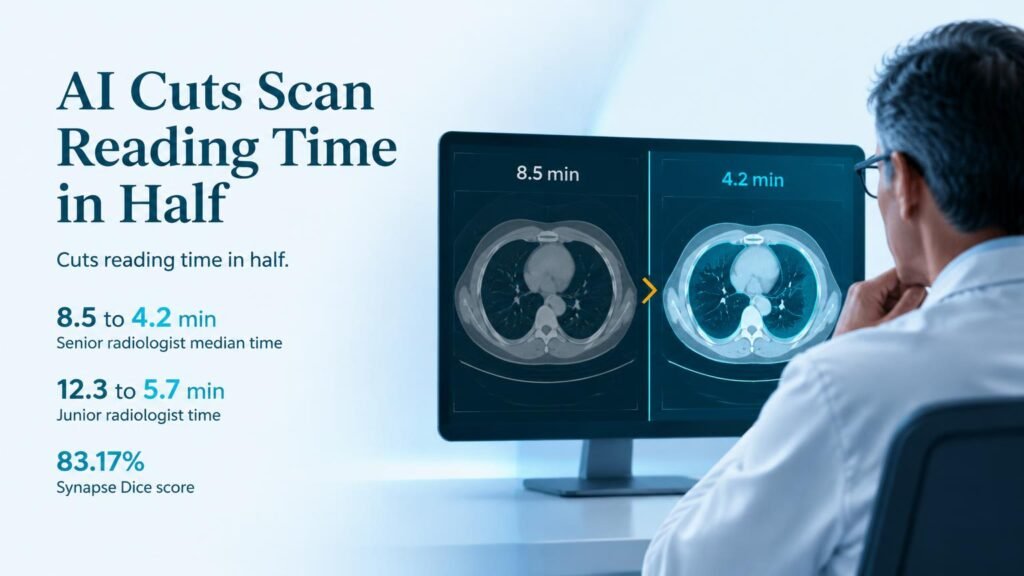
Evaluated on the public Synapse dataset, AMIS-Net recorded an overall Dice similarity coefficient of 83.17% and a 95th percentile Hausdorff Distance of 20.89 mm. Across specific organs, performance varied from a low Dice score of 74.85% for the esophagus to a high of 94.21% for the liver. The architecture consistently outperformed U-Net, ResUNet, and STUNet across the CHAOS dataset, Synapse, and a proprietary clinical dataset.
- Senior radiologist reading times dropped from 8.5 to 4.2 minutes per case.
- Junior radiologist reading times dropped from 12.3 to 5.7 minutes per case.
- Per-organ segmentation accuracy ranged from 74.85% for the esophagus to 94.21% for the liver.
- Outperformed standard U-Net, ResUNet, and STUNet models across benchmark and clinical validation datasets.
Clinical Impact and Practical Limitations
When deployed in a clinical workflow for liver tumors and intracranial hemorrhage, the tool dramatically reduced manual labor. Senior radiologists cut their median reading time from 8.5 to 4.2 minutes, while junior radiologists reduced theirs from 12.3 to 5.7 minutes. The system also lowered missed diagnosis rates and improved overall diagnostic accuracy across reader experience levels.
However, time savings in a controlled reader study do not prove that automated segmentation improves final patient outcomes. The performance drop on narrow, highly variable structures like the esophagus (74.85% Dice) shows that manual corrections remain necessary for complex anatomy. Clinical administrators should view these tools as efficiency aids to reduce fatigue, not as drop-in replacements for human verification.
Read the full paper in Scientific Reports.