A new deep learning model closes the diagnostic gap between junior radiologists and seasoned specialists by analyzing ultrasound videos instead of static images.
Static ultrasound images often fail to capture the complex blood flow and tissue movement needed to accurately diagnose swollen lymph nodes. This diagnostic uncertainty forces patients into unnecessary, painful biopsies or delays critical cancer treatment. A multicenter study suggests that the solution lies in analyzing dynamic ultrasound videos rather than frozen frames.
This shift from static images to dual-modality video analysis represents a practical leap for clinical workflows. Static imaging forces clinicians to rely on subjective interpretations of shape and size. In contrast, dual-modality video tracks the continuous flow of blood, which reveals the chaotic vessel branching typical of malignant tumors. This objective data stream helps eliminate the cognitive bias that often leads to diagnostic errors, allowing junior clinicians to perform at near-expert levels.
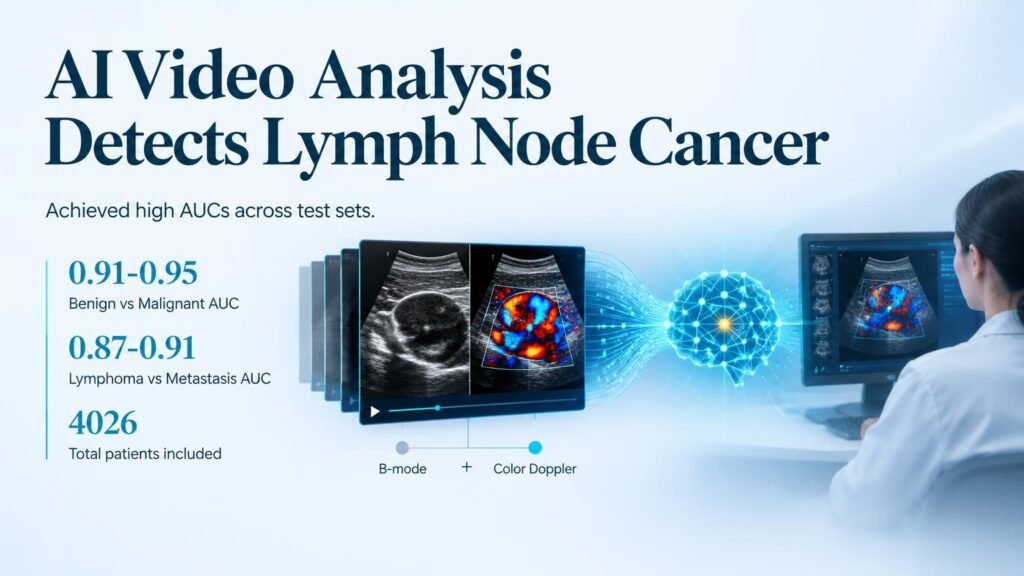
The study evaluated 4,026 patients (including 2,175 males) with a median age of 52.6 years across five clinical centers between June 2019 and December 2025. Researchers trained the model, named DMUVL-DiagNet, on a retrospective dataset of 1,616 video cases. They then validated its performance using an external prospective dataset of 454 videos and a static image set of 1,956 cases.
Key diagnostic metrics
- The model achieved an AUC of 0.91 to 0.95 for distinguishing benign from malignant lymph nodes.
- It reached an AUC of 0.87 to 0.91 when differentiating lymphoma from metastatic cancer.
- With AI assistance, junior radiologists improved their AUC from 0.72 to 0.88 in the benign-malignant task.
- Junior readers also saw their lymphoma-metastasis diagnostic AUC jump from 0.58 to 0.77.
Closing the expertise gap
The real value here is the dramatic improvement in junior radiologist performance. A jump from an AUC of 0.58 to 0.77 for differentiating lymphoma from metastasis moves a junior clinician from near-random guessing to a highly reliable diagnostic standard. This leveling of expertise could significantly reduce referral delays and prevent misdiagnoses in community clinics lacking senior specialists.
However, diagnostic accuracy in a study setting does not automatically translate to improved patient survival or lower healthcare costs. While the model showed strong generalizability across multiple centers, the training relied on retrospective data, which can introduce selection bias. We must also note that the model was tested on a static image set of 1,956 cases to prove its versatility, yet its peak performance remains tied to video inputs. Implementing video-based AI in clinics requires standardized video acquisition protocols, as slight hand movements by the technician can alter the video feed. Until these workflow hurdles are cleared, the tool remains a promising assistant rather than a replacement for human oversight.
Read the full study in Radiology: Artificial Intelligence.