AI spots esophageal cancer on routine chest scans
An AI model trained to find esophageal cancer on routine chest scans could soon turn standard lung screenings into a dual-detection system for early-stage malignancies.
Clinicians have long ruled out noncontrast CT scans for esophageal cancer screening because the collapsed, shifting tissue of the esophagus hides early tumors. A new study challenges this clinical assumption by using AI to analyze standard chest scans already sitting in hospital databases. By piggybacking on routine lung cancer screenings, this approach could identify high-risk patients before they ever show symptoms.
That shift in workflow is where the real value lies.
Instead of forcing patients to undergo invasive endoscopies, health systems can use existing imaging infrastructure to flag high-risk individuals. Researchers built the EAGLE model using data from 6,813 patients across two clinical centers. They then validated the tool across 12 centers in three countries, analyzing a massive dataset of 80,612 patients. The testing spanned opportunistic screening of existing scans, low-dose CT programs, and prospective hospital workflows.
High specificity, lower sensitivity
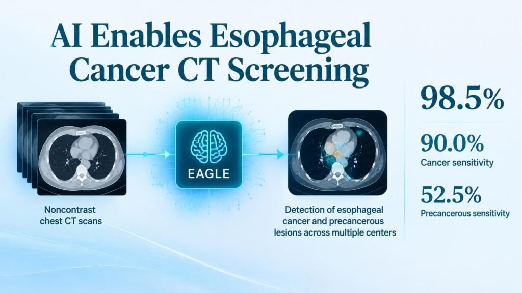
The AI proved highly capable of ruling out healthy patients, which is critical for preventing unnecessary, invasive follow-up endoscopies. In the opportunistic screening cohort of 11,466 patients, the model achieved a specificity of 98.5%. However, its ability to catch early-stage disease was mixed, showing that the tool is not a magic bullet for all stages of progression.
- Caught 90.0% of active esophageal cancers in the opportunistic screening cohort.
- Detected only 52.5% of precancerous lesions in the same group.
- Achieved a 42.2% positive predictive value in prospective validation of 17,446 patients.
- Reduced false-positive rates by 72.7% in a real-world calibration cohort of 35,402 patients.
Low-dose CT validation across 1,607 patients showed comparable performance, confirming that the model works on lower-resolution scans. Furthermore, in a real-world low-dose screening cohort of 10,959 patients, the specificity reached an outstanding 99.94%.
The screening bottleneck
These numbers show a clear limitation: the system is far better at spotting fully formed cancers than the precancerous lesions that are most treatable. In a paired CT-endoscopy cohort of 702 patients, sensitivity for precancerous lesions rose to 65.0% only when researchers adjusted the model to a higher-sensitivity operating point. This trade-off means clinicians must decide whether they want to minimize false alarms or maximize early detection.
This is not a replacement for endoscopy, which remains the gold standard. Instead, the practical takeaway is operational triaging. By running this AI silently in the background of routine lung cancer screenings, hospitals can flag a subset of high-risk patients for targeted endoscopic referrals, making population-level screening logistically viable for the first time.
Read the full study in Nature Medicine.