Adding radiology text to visual AI models stops them from failing when hyperparameters change.
Can we trust an AI that classifies brain tumors if a tiny tweak to its learning rate tanks its accuracy by half? That is the quiet crisis in medical imaging. Most deep learning models are hyper-sensitive black boxes that perform beautifully in a lab but fail under slight environmental shifts.
A new preprint introducing TumorCLIP exposes this vulnerability. The researchers ran a comprehensive unimodal benchmark across eight visual backbones, including EfficientNet-B0, MobileNetV3-Large, ResNet50, DenseNet121, ViT, DeiT, Swin Transformer, and MambaOut. They found performance swings exceeding 60 percentage points based purely on optimizer and learning-rate choices.
That volatility is terrifying for clinical deployment.
The hidden fragility of AI
To find a stable foundation, the team tested various architectures. DenseNet121 emerged as the most resilient, showing a 97.6% stability-accuracy trade-off within the evaluated grid. While other studies focus on complex new architectures like SwinBTS for 3D segmentation, this finding suggests that older, convolutional backbones might actually be safer starting points for clinical use because they are less erratic.
But stability alone is not enough. Purely visual models lack semantic reasoning. They classify pixels without understanding what a radiologist actually looks for in an MRI scan.
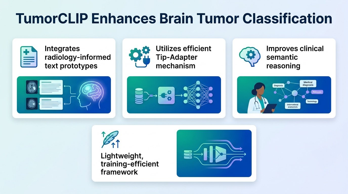
Anchoring pixels to radiology text
TumorCLIP solves this by fusing the stable DenseNet121 visual encoder with frozen CLIP-derived text prototypes. It uses a lightweight Tip-Adapter mechanism to align image features with actual radiological concepts. Instead of learning from scratch, the AI is guided by pre-defined clinical descriptions.
This hybrid approach, similar to the multi-modal strategies explored in recent LLM-based diagnosis systems, yields impressive metrics:
- Overall test accuracy reached 98.5%, outperforming the unimodal baseline.
- The positive semantic margin rate jumped to 96.9%, compared to just 42.1% for the baseline.
- The model showed significantly reduced degradation when tested on entirely different datasets, especially for highly variable tumors like glioma.
Why clinical context wins
This is not just about a minor accuracy bump. The real value is the semantic margin rate. A 96.9% rate means the model’s internal decision-making aligns with actual medical concepts, making its predictions interpretable to human doctors. It reduces inter-class misclassification by grounding visual patterns in medical language.
There are limitations to keep in mind. The framework relies on frozen text prototypes, meaning its vocabulary is fixed and cannot adapt dynamically during training. It was also validated within specific training protocols, meaning real-world clinical environments might still present unforeseen edge cases.
Even so, the lesson is clear. The path to reliable medical AI is not about building larger, more complex visual networks. It is about anchoring existing visual models to the structured language of medicine.
Read the full preprint on medRxiv.