⚡ Quick Summary
This study utilized multimodal contrastive learning on resting-state functional MRI (rs-fMRI) data from 56 patients to assess the recovery of whole-brain networks following hypothalamic hamartoma surgery. The results demonstrated a mean accuracy of 0.85 to 0.90 in distinguishing pre-operative from post-operative brain states across various networks.
🔍 Key Details
- 📊 Dataset: 56 patients with hypothalamic hamartoma
- 🧩 Features used: rs-fMRI data, including 3D spatial and 1D temporal ICA maps
- ⚙️ Technology: Two-stage contrastive learning algorithm
- 🏆 Performance: Mean accuracy: 0.85 to 0.90, Sensitivity: 0.79 to 0.90, Specificity: 0.87 to 0.93
🔑 Key Takeaways
- 🧠 Contrastive learning effectively detects functional shifts in critical cortical networks.
- 📈 High accuracy achieved in classifying brain states pre- and post-surgery.
- 🔍 Visualization techniques like t-SNE revealed clear separations in network states.
- 💡 Findings inform understanding of whole-brain network functioning post-surgery.
- 🌟 Future studies are encouraged to compare HH patients with healthy controls.
📚 Background
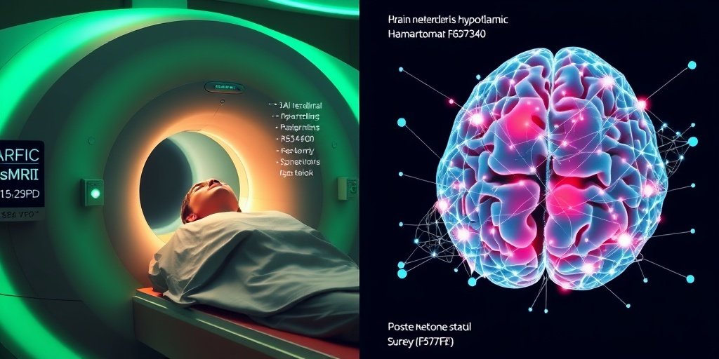
Hypothalamic hamartoma (HH) is a rare brain tumor that can lead to severe epilepsy and cognitive impairment. Traditional surgical interventions aim to alleviate seizures, but the broader impact on brain networks has not been thoroughly quantified. Understanding these changes is crucial for assessing the effectiveness of surgical treatments and improving patient outcomes.
🗒️ Study
This retrospective study analyzed rs-fMRI data from 56 patients diagnosed with hypothalamic hamartoma. The researchers developed a novel two-stage contrastive learning algorithm to classify the brain’s motor, vision, language, frontal, and temporal networks before and six months after surgery. This approach aimed to uncover subtle changes in brain connectivity that conventional analyses might miss.
📈 Results
The study’s findings revealed a mean accuracy ranging from 0.85 to 0.90 in distinguishing pre-operative from post-operative states across the five networks analyzed. Sensitivity and specificity metrics were also impressive, with sensitivity values between 0.79 and 0.90 and specificity values from 0.87 to 0.93. The area under the curve (AUC) ranged from 0.90 to 0.94, indicating robust performance in the classification task.
🌍 Impact and Implications
The implications of this study are significant for both clinical practice and future research. By employing contrastive learning, researchers can gain deeper insights into the functional recovery of brain networks after surgery. This methodology not only enhances our understanding of the effects of hypothalamic hamartoma surgery but also sets the stage for future studies comparing HH patients to healthy controls, ultimately aiming to quantify network recovery more effectively.
🔮 Conclusion
This research highlights the potential of contrastive learning in neuroimaging, particularly in assessing functional changes in the brain following surgical interventions. The ability to detect subtle shifts in brain networks can lead to improved patient management and outcomes. As we move forward, further exploration in this area promises to enrich our understanding of brain recovery processes and enhance therapeutic strategies.
💬 Your comments
What are your thoughts on the use of advanced machine learning techniques in neuroimaging? We would love to hear your insights! 💬 Leave your comments below or connect with us on social media:
Multimodal contrastive learning on rs-fMRI to quantify whole-brain network recovery after hypothalamic hamartoma surgery.
Abstract
INTRODUCTION: Epilepsy due to hypothalamic hamartoma (HH) is associated with epileptic encephalopathy and often requires surgical intervention, as medications are ineffective at reducing the seizures. However, the first step of disentangling the impact of the surgery on the broader whole-brain networks, a biomarker of encephalopathy compared to controls, is not quantified. Subtle pre- and post-operative networks can elude conventional rs-fMRI analysis.
METHODS: We retrospectively analyzed rs-fMRI from 56 HH patients scanned before and 6 months after surgery. We developed a two-stage contrastive learning-based algorithm to classify the motor, vision, language, frontal, and temporal networks as pre- vs post-operative. In stage one, a multimodal contrastive encoder jointly ingests 3D spatial Independent Component Analysis (ICA) maps and their corresponding 1D temporal ICA time series to learn embeddings that distinguish pre-operative from post-operative states for each network while separating embeddings of different networks. In stage two, a lightweight classifier refines these embeddings, augmented by original ICA inputs, to classify each network as pre-operative or post-operative.
RESULTS: Visualization of the learned feature space with t-SNE revealed clear separation by pre- vs post-surgical condition across all five networks. Across networks, mean accuracy ranged from 0.85 to 0.90, sensitivity from 0.79 to 0.90, specificity from 0.87 to 0.93, F1-score from 0.83 to 0.90 and AUC from 0.90 to 0.94 in stratified cross validation.
CONCLUSIONS: Contrastive learning can sensitively detect functional shifts in critical cortical networks that previous traditional analyses may overlook. These findings inform broader shifts in whole-brain network functioning following effective HH surgery and establish a featurewise distinction between preoperative and postoperative states, motivating future studies that compare HH patients to healthy controls to quantify network recovery.
Author: [‘Jeyabose A’, ‘Robinson B’, ‘Boerwinkle VL’, ‘Leggio O’, ‘Kazemi MH’]
Journal: Biomed Eng Online
Citation: Jeyabose A, et al. Multimodal contrastive learning on rs-fMRI to quantify whole-brain network recovery after hypothalamic hamartoma surgery. Multimodal contrastive learning on rs-fMRI to quantify whole-brain network recovery after hypothalamic hamartoma surgery. 2025; 24:125. doi: 10.1186/s12938-025-01458-6