Hospital buyers assume all top-tier radiology AI performs the same, but new head-to-head data reveals critical trade-offs that could compromise patient care if ignored.
If every commercial AI claims near-perfect accuracy for spotting brain bleeds, how do you choose the right one? Most hospitals buy AI based on a single, high-level metric like sensitivity. This is a mistake. A new study comparing four commercial algorithms in a real-world emergency room shows that identical headline accuracy hides massive differences in how these tools actually behave.
This challenges the industry habit of treating clinical AI as an interchangeable commodity. The findings suggest that procurement teams must stop asking if an AI works, and start asking how its specific errors align with their clinical workflow.
The Hidden Performance Gap
The trial evaluated **436** non-contrast brain CT scans from an emergency department, representing **209** male patients and a cohort with a mean age of **62** years. Three neuroradiologists established the ground truth. While previous research has shown that deep learning boosts radiologist detection of intracranial hemorrhage, as documented in a Cureus study, this head-to-head trial looked deeper at the math behind the tools.
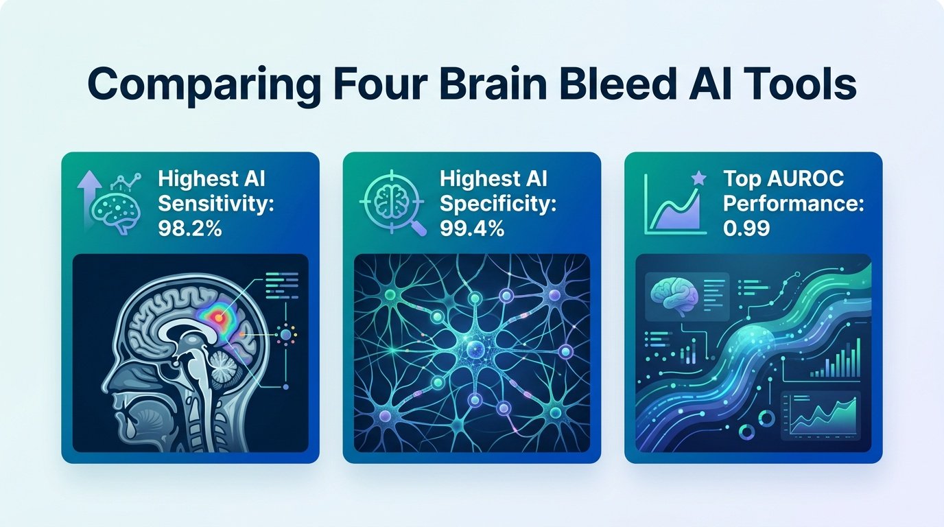
On the surface, the algorithms looked identical. All four solutions achieved high AUROC scores ranging from **0.96 to 0.99** and sensitivity from **0.85 to 0.92**. If you only looked at these baseline metrics, you would buy the cheapest option.
But the secondary metrics told a completely different story. Solution B stood out with the highest AUPRC of **0.98** and the lowest Brier score of **0.02**. In binary performance, Solutions B and D showed significantly higher specificity (**1.00 and 0.99**), precision (**0.90 to 0.98**), and F1 scores (**0.87 to 0.94**) than their competitors.
Volume Tracking and Workflow Realities
The differences became even sharper when measuring the actual volume of the bleeds. Solution D excelled here, showing the lowest mean volumetric difference of **-0.87 mm³** and the narrowest limits of agreement of **-13.4 to 11.6** relative to the neuroradiologists. This matters because measuring bleed volume is crucial for tracking patient decline, a point emphasized in earlier automated segmentation research.
This means a hospital must choose its poison. If your ER is drowning in false alarms, you need Solution B or D to keep specificity high and reduce alert fatigue. But if your neurosurgeons rely on precise volume tracking to plan surgeries, Solution D is the clear winner.
Key Performance Metrics
- All four solutions achieved high AUROC scores between **0.96 and 0.99**.
- Solutions B and D achieved superior specificity of **1.00 and 0.99** respectively.
- Solution D offered the most precise volume tracking with a mean difference of **-0.87 mm³**.
Real-World Limitations
We must be honest about the limits of this data. This was a single-center study over a short two-month window in 2024. The sample size of **436** scans, while valuable, may not reflect patient diversity or different scanner hardware in other hospital networks.
Ultimately, this study proves that “one size fits all” is a myth in medical AI. Hospitals must run their own multimetric evaluations to match an algorithm’s unique strengths to their specific clinical bottlenecks.
Read the full study in Diagnostic and Interventional Radiology.