⚡ Quick Summary
This study developed an AI-driven radiomics prediction model to differentiate between benign and malignant pulmonary nodules with a 3D consolidation-to-tumor ratio (CTR) ≥ 50%. The model demonstrated moderate accuracy, with an AUC of 0.757 in the validation set, highlighting its potential in clinical diagnostics.
🔍 Key Details
- 📊 Dataset: 370 pulmonary nodules (78 benign, 292 malignant)
- 🧩 Features used: 108 radiomic features
- ⚙️ Technology: Lasso regression and logistic regression
- 🏆 Performance: Training set AUC 0.721, Validation set AUC 0.757
🔑 Key Takeaways
- 🔬 AI-driven radiomics can enhance diagnostic accuracy for pulmonary nodules.
- 📈 The model identified three significant factors: patient age, solid component volume, and mean CT value.
- 🧪 The study utilized data from five medical centers, ensuring a diverse dataset.
- 📊 The model’s AUC values indicate moderate predictive ability and stability.
- 🌟 Future research could focus on improving the model’s precision and clinical applicability.
- 🏥 Clinical relevance is significant, especially for nodules with a CTR ≥ 50%.
- 📅 Study published in BMC Medical Imaging in 2025.
📚 Background
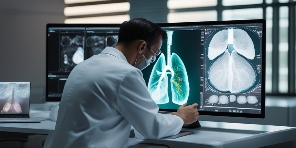
Diagnosing the nature of pulmonary nodules, particularly those with a 3D CTR ≥ 50%, poses a significant challenge in clinical practice. Malignant nodules are often more invasive, making accurate differentiation crucial for patient management. The integration of artificial intelligence and radiomics analysis offers a promising avenue to enhance diagnostic accuracy and improve patient outcomes.
🗒️ Study
The study involved a comprehensive analysis of 2,591 pulmonary nodules collected from five medical centers, ultimately focusing on 370 nodules that met the inclusion criteria. Researchers employed Lasso regression with 10-fold cross-validation to filter features, followed by logistic regression to construct the prediction model. The efficacy of the model was evaluated using ROC, DCA curves, and calibration plots.
📈 Results
The Lasso regression identified 18 significant features from the initial 108, leading to the formulation of a logistic regression equation. The model achieved an AUC of 0.721 in the training set and 0.757 in the validation set, indicating its potential for reliable differentiation between benign and malignant nodules.
🌍 Impact and Implications
The findings of this study could significantly impact clinical practice by providing a reliable tool for the assessment of pulmonary nodules. The ability to accurately differentiate between benign and malignant nodules can lead to better patient management and treatment decisions, ultimately improving patient outcomes. As AI technology continues to evolve, its integration into diagnostic processes holds great promise for enhancing healthcare delivery.
🔮 Conclusion
This study highlights the potential of AI-driven radiomics in improving the diagnostic accuracy of pulmonary nodules with a CTR ≥ 50%. While the model shows moderate accuracy, further research is essential to refine its precision and clinical utility. The future of AI in healthcare looks promising, and continued exploration in this field is encouraged!
💬 Your comments
What are your thoughts on the use of AI in diagnosing pulmonary nodules? We would love to hear your insights! 💬 Leave your comments below or connect with us on social media:
Development of a clinical prediction model for benign and malignant pulmonary nodules with a CTR ≥ 50% utilizing artificial intelligence-driven radiomics analysis.
Abstract
OBJECTIVE: In clinical practice, diagnosing the benignity and malignancy of solid-component-predominant pulmonary nodules is challenging, especially when 3D consolidation-to-tumor ratio (CTR) ≥ 50%, as malignant ones are more invasive. This study aims to develop and validate an AI-driven radiomics prediction model for such nodules to enhance diagnostic accuracy.
METHODS: Data of 2,591 pulmonary nodules from five medical centers (Zhengzhou People’s Hospital, etc.) were collected. Applying exclusion criteria, 370 nodules (78 benign, 292 malignant) with 3D CTR ≥ 50% were selected and randomly split 7:3 into training and validation cohorts. Using R programming, Lasso regression with 10-fold cross-validation filtered features, followed by univariate and multivariate logistic regression to construct the model. Its efficacy was evaluated by ROC, DCA curves and calibration plots.
RESULTS: Lasso regression picked 18 non-zero coefficients from 108 features. Three significant factors-patient age, solid component volume and mean CT value-were identified. The logistic regression equation was formulated. In the training set, the ROC AUC was 0.721 (95%CI: 0.642-0.801); in the validation set, AUC was 0.757 (95%CI: 0.632-0.881), showing the model’s stability and predictive ability.
CONCLUSION: The model has moderate accuracy in differentiating benign from malignant 3D CTR ≥ 50% nodules, holding clinical potential. Future efforts could explore more to improve its precision and value.
CLINICAL TRIAL NUMBER: Not applicable.
Author: [‘Shi W’, ‘Hu Y’, ‘Chang G’, ‘Qian H’, ‘Yang Y’, ‘Song Y’, ‘Wei Z’, ‘Gao L’, ‘Yi H’, ‘Wu S’, ‘Wang K’, ‘Huo H’, ‘Wang S’, ‘Mao Y’, ‘Ai S’, ‘Zhao L’, ‘Li X’, ‘Zheng H’]
Journal: BMC Med Imaging
Citation: Shi W, et al. Development of a clinical prediction model for benign and malignant pulmonary nodules with a CTR ≥ 50% utilizing artificial intelligence-driven radiomics analysis. Development of a clinical prediction model for benign and malignant pulmonary nodules with a CTR ≥ 50% utilizing artificial intelligence-driven radiomics analysis. 2025; 25:21. doi: 10.1186/s12880-024-01533-9