⚡ Quick Summary
This study presents a novel method for the automated detection and segmentation of breast cancer focal lesions in ultrasound images, achieving an impressive 91.89% detection rate and an IoU accuracy of 0.871. The proposed algorithm enhances the diagnostic capabilities of ultrasound imaging, addressing challenges posed by noise and artifacts.
🔍 Key Details
- 📊 Dataset: 52 ultrasound videos with histologically proven suspicious lesions
- ⚙️ Technology: Random forest classifier for region of interest detection
- 🔍 Methodology: Two-stage image processing for lesion segmentation
- 🏆 Performance: 91.89% average frequency of lesion detection per frame
- 📏 Accuracy: IoU metric of 0.871 for contour selection
🔑 Key Takeaways
- 🖼️ Ultrasound imaging is crucial for breast cancer diagnosis but often affected by noise.
- 🤖 Deep learning methods face challenges in model substantiation and training data collection.
- 🔬 The proposed method utilizes a random forest classifier for effective lesion detection.
- 📈 High detection rates indicate the method’s potential for clinical application.
- 🌟 The study contributes to the ongoing development of automated diagnostic tools in oncology.
- 📅 Published in: Sensors (Basel), 2025.
- 🆔 PMID: 40096452.
📚 Background
Breast cancer remains one of the leading causes of cancer-related mortality among women worldwide. Early detection is critical for improving patient outcomes, and ultrasound imaging plays a vital role in the differential diagnosis of breast lesions. However, the quality of ultrasound images can be compromised by various artifacts and noise, making accurate analysis challenging. This study aims to enhance the diagnostic process through advanced image processing techniques.
🗒️ Study
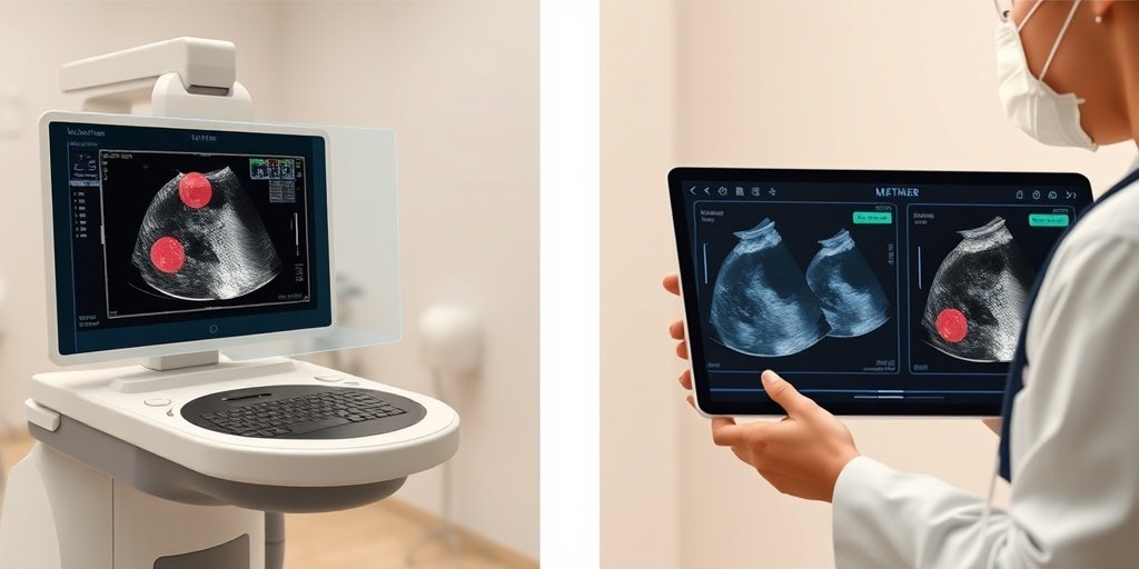
Conducted by a team of researchers, this study focuses on developing an automated method for detecting and segmenting pathological lesions in breast ultrasound images. The methodology consists of two main stages: first, identifying regions of interest using a random forest classifier, and second, delineating the contours of lesions based on pixel brightness differences. The study utilized a dataset of 52 ultrasound videos containing confirmed suspicious lesions for testing.
📈 Results
The results of the study were promising, with an average frequency of lesion detection per frame reaching 91.89%. Additionally, the accuracy of contour selection, measured by the Intersection over Union (IoU) metric, was found to be 0.871. These metrics indicate that the proposed method is highly effective in segmenting suspicious lesions, which could significantly aid radiologists in their diagnostic efforts.
🌍 Impact and Implications
The implications of this study are substantial for the field of medical imaging and oncology. By improving the accuracy and efficiency of breast cancer diagnosis through automated segmentation techniques, healthcare providers can enhance patient care and potentially improve survival rates. This research paves the way for further advancements in the integration of artificial intelligence in medical diagnostics, ultimately leading to better healthcare outcomes.
🔮 Conclusion
This study highlights the transformative potential of automated image processing techniques in the realm of breast cancer diagnosis. With a high detection rate and accuracy in lesion segmentation, the proposed method represents a significant step forward in utilizing technology to support healthcare professionals. Continued research and development in this area could lead to even more sophisticated diagnostic tools, improving the overall quality of care for patients.
💬 Your comments
What are your thoughts on the integration of automated segmentation methods in breast cancer diagnostics? We would love to hear your insights! 💬 Share your comments below or connect with us on social media:
Automated Segmentation of Breast Cancer Focal Lesions on Ultrasound Images.
Abstract
Ultrasound (US) remains the main modality for the differential diagnosis of changes revealed by mammography. However, the US images themselves are subject to various types of noise and artifacts from reflections, which can worsen the quality of their analysis. Deep learning methods have a number of disadvantages, including the often insufficient substantiation of the model, and the complexity of collecting a representative training database. Therefore, it is necessary to develop effective algorithms for the segmentation, classification, and analysis of US images. The aim of the work is to develop a method for the automated detection of pathological lesions in breast US images and their segmentation. A method is proposed that includes two stages of video image processing: (1) searching for a region of interest using a random forest classifier, which classifies normal tissues, (2) selecting the contour of the lesion based on the difference in brightness of image pixels. The test set included 52 ultrasound videos which contained histologically proven suspicious lesions. The average frequency of lesion detection per frame was 91.89%, and the average accuracy of contour selection according to the IoU metric was 0.871. The proposed method can be used to segment a suspicious lesion.
Author: [‘Pasynkov D’, ‘Egoshin I’, ‘Kolchev A’, ‘Kliouchkin I’, ‘Pasynkova O’, ‘Saad Z’, ‘Daou A’, ‘Abuzenar EM’]
Journal: Sensors (Basel)
Citation: Pasynkov D, et al. Automated Segmentation of Breast Cancer Focal Lesions on Ultrasound Images. Automated Segmentation of Breast Cancer Focal Lesions on Ultrasound Images. 2025; 25:(unknown pages). doi: 10.3390/s25051593
2 Comments
Weekly Health & AI Digest – March 20, 2025 - Yesil Science
[…] Automated Segmentation of Breast Cancer Focal Lesions on Ultrasound Images. […]
Weekly Health & AI Digest – March 21, 2025 - Yesil Science
[…] 🔹 Automated Segmentation of Breast Cancer Focal Lesions on Ultrasound Images. […]