⚡ Quick Summary
This study utilized AI foundation models to analyze histopathologic features in HPV-positive head and neck squamous cell carcinomas (HNSCCs), revealing distinctive histological characteristics and achieving a validation sensitivity of 81% and specificity of 92% for HPV detection.
🔍 Key Details
- 📊 Dataset: 981 HNSCC patients from public and institutional datasets
- 🧩 Features used: Hematoxylin and eosin (H&E) stained slides
- ⚙️ Technology: UNI (self-supervised learning) and HistoXGAN (generative adversarial network)
- 🏆 Performance: Sensitivity 0.81, Specificity 0.92
🔑 Key Takeaways
- 🔬 AI foundation models can effectively decode the histopathologic landscape of HPV-positive HNSCCs.
- 🖼️ Distinctive features of HPV-positive histology include smaller, paler nuclei and indistinct cell borders.
- 📈 Accurate prediction of HPV status can be achieved directly from histology images.
- 🌟 Three overlapping subtypes of HPV-positive histology were identified: border, inflamed, and stroma.
- 💡 This approach offers a valuable tool for low-resource clinical settings.
📚 Background
The influence of human papillomavirus (HPV) on the pathobiology of HNSCCs is well-documented, yet the specific histologic features that correlate with HPV presence have remained unclear. Traditional methods of detecting HPV often rely on subjective assessments, which can lead to variability in diagnosis. The integration of artificial intelligence into this field presents an opportunity to enhance diagnostic accuracy and objectivity.
🗒️ Study
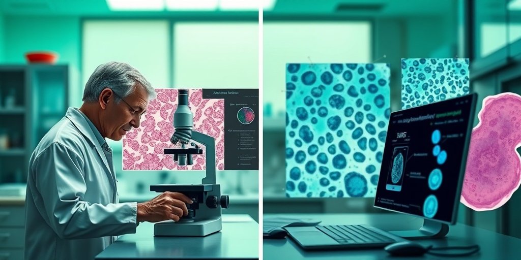
This study analyzed H&E images from 981 patients diagnosed with HNSCC. Researchers employed the UNI foundation model based on self-supervised learning to map the histological landscape and identify features that differentiate HPV-positive from HPV-negative tumors. Additionally, they utilized HistoXGAN to generate synthetic histology images for further analysis by pathologists.
📈 Results
The analysis of AI-generated synthetic images revealed distinctive histological features associated with HPV positivity. These included smaller, paler nuclei, purpler cytoplasm, and rounded tumor contours. The identified SSL feature axes allowed for an impressive prediction accuracy of HPV status, with a sensitivity of 81% and specificity of 92%. Furthermore, the study categorized HPV-positive histology into three subtypes, enhancing our understanding of the disease’s variability.
🌍 Impact and Implications
The findings from this study have significant implications for the field of oncology, particularly in the diagnosis and management of HPV-positive HNSCCs. By leveraging AI technologies, healthcare providers can achieve more accurate and explainable detection of HPV directly from histology, which is especially beneficial in low-resource clinical settings. This advancement could lead to improved patient outcomes and more tailored treatment strategies.
🔮 Conclusion
This research highlights the transformative potential of AI in pathology, particularly in decoding the complex histopathologic landscape of HPV-positive HNSCCs. The ability to accurately detect HPV presence from histology images not only enhances diagnostic capabilities but also paves the way for future innovations in cancer care. Continued exploration in this area is essential for advancing our understanding and treatment of HPV-related cancers.
💬 Your comments
What are your thoughts on the integration of AI in pathology? Do you believe it can significantly change cancer diagnostics? 💬 Share your insights in the comments below or connect with us on social media:
Analysis of AI foundation model features decodes the histopathologic landscape of HPV-positive head and neck squamous cell carcinomas.
Abstract
OBJECTIVES: Human papillomavirus (HPV) influences the pathobiology of Head and Neck Squamous Cell Carcinomas (HSNCCs). While deep learning shows promise in detecting HPV from hematoxylin and eosin (H&E) stained slides, the histologic features utilized remain unclear. This study leverages artificial intelligence (AI) foundation models to characterize histopathologic features associated with HPV presence and objectively describe patterns of variability in the HPV-positive space.
MATERIALS AND METHODS: H&E images from 981 HNSCC patients across public and institutional datasets were analyzed. We used UNI, a foundation model based on self-supervised learning (SSL), to map the landscape of HNSCC histology and identify the axes of SSL features that best separate HPV-positive and HPV-negative tumors. To interpret the histologic features that vary across different regions of this landscape, we used HistoXGAN, a pretrained generative adversarial network (GAN), to generate synthetic histology images from SSL features, which a pathologist rigorously assessed.
RESULTS: Analyzing AI-generated synthetic images found distinctive features of HPV-positive histology, such as smaller, paler, more monomorphic nuclei; purpler, amphophilic cytoplasm; and indistinct cell borders with rounded tumor contours. The SSL feature axes we identified enabled accurate prediction of HPV status from histology, achieving validation sensitivity and specificity of 0.81 and 0.92, respectively. Our analysis subdivided image tiles from HPV-positive histology into three overlapping subtypes: border, inflamed, and stroma.
CONCLUSION: Foundation-model-derived synthetic pathology images effectively capture HPV-related histology. Our analysis identifies distinct subtypes within HPV-positive HNSCCs and enables accurate, explainable detection of HPV presence directly from histology, offering a valuable approach for low-resource clinical settings.
Author: [‘Hieromnimon HM’, ‘Trzcinska A’, ‘Wen FT’, ‘Howard FM’, ‘Dolezal JM’, ‘Dyer E’, ‘Kochanny S’, ‘Schulte JJ’, ‘Wang C’, ‘Chen H’, ‘Chin J’, ‘Blair E’, ‘Agrawal N’, ‘Rosenberg A’, ‘Vokes E’, ‘Katipally R’, ‘Juloori A’, ‘Izumchenko E’, ‘Lingen MW’, ‘Cipriani N’, ‘Jalaly JB’, ‘Basu D’, ‘Riesenfeld SJ’, ‘Pearson AT’]
Journal: Oral Oncol
Citation: Hieromnimon HM, et al. Analysis of AI foundation model features decodes the histopathologic landscape of HPV-positive head and neck squamous cell carcinomas. Analysis of AI foundation model features decodes the histopathologic landscape of HPV-positive head and neck squamous cell carcinomas. 2025; 163:107207. doi: 10.1016/j.oraloncology.2025.107207