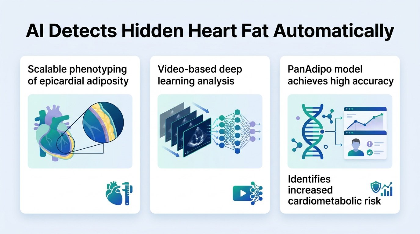
A new AI model extracts metabolic risk data directly from routine heart ultrasounds, bypassing the need for expensive CT scans.
Doctors routinely use echocardiograms to check how well a heart pumps blood. But what if they are ignoring a massive warning sign hidden in plain sight on the very same screens?
The fat surrounding the heart, known as epicardial adipose tissue, is a major driver of cardiovascular and metabolic disease. Yet measuring it usually requires a costly, radiation-heavy cardiac CT scan. This diagnostic barrier leaves a critical cardiovascular risk factor unmeasured for the vast majority of patients.
This study challenges the idea that echocardiograms are only good for structural and plumbing checks. By training AI to see what human eyes overlook, clinicians can turn a standard, cheap test into a metabolic warning system. This matters because body mass index is a notoriously blunt tool. Patients can have a normal weight but carry dangerous fat around their organs. This tool decouples metabolic risk from body size.
Finding the hidden fat
Researchers built PanAdipo, a deep learning model trained on 1,114,441 ultrasound videos from 28,797 patients. The system learned to recognize prominent heart fat using expert annotations from a fraction of those studies. The team then validated the model across diverse clinical environments.
To prove the model worked, the researchers tested it across four distinct groups. These included an emergency department cohort of 10,957 patients and the community-based MESA study of 2,740 participants. The results were remarkably consistent. In the main test set, PanAdipo flagged prominent heart fat with an AUROC of 0.91, easily beating traditional measures of cardiac structure.
Beyond body mass index
The real value of this tool lies in its independence from body weight. The AI’s fat scores showed only modest correlation with BMI, with a Spearman’s rho of just 0.19 to 0.40. This proves the tool measures a distinct biological signal rather than just reflecting a patient’s overall weight.
When compared to gold-standard CT scans in 5,594 patients, the AI’s ultrasound-based score correlated strongly with actual heart fat, yielding a Spearman’s rho of 0.75. Key findings from the clinical validation include:
- An independent link to higher insulin resistance and triglycerides, even in patients with normal blood sugar.
- A 1.25 hazard ratio for newly documented metabolic diseases, including liver diseases like MASLD and MASH, for every one standard deviation increase in the AI score.
- Strong spatial accuracy, with explainability maps showing the AI focused precisely on the epicardial areas throughout the entire cardiac cycle.
The clinical catch
Despite the strong data, implementation faces hurdles. The model relies on retrospective data from specific health systems, and clinical workflows are already crowded. Adding another AI marker to an ultrasound report does not automatically translate to better patient care.
Furthermore, knowing a patient has high heart fat is only useful if clinicians have clear pathways to treat it. Without integrated care plans, this tool risks generating data points that doctors do not know how to act upon. Still, this approach shifts the paradigm of cardiac imaging. It proves that existing diagnostic hardware holds vast pools of unused, clinically valuable data.
Read the full preprint in medRxiv.