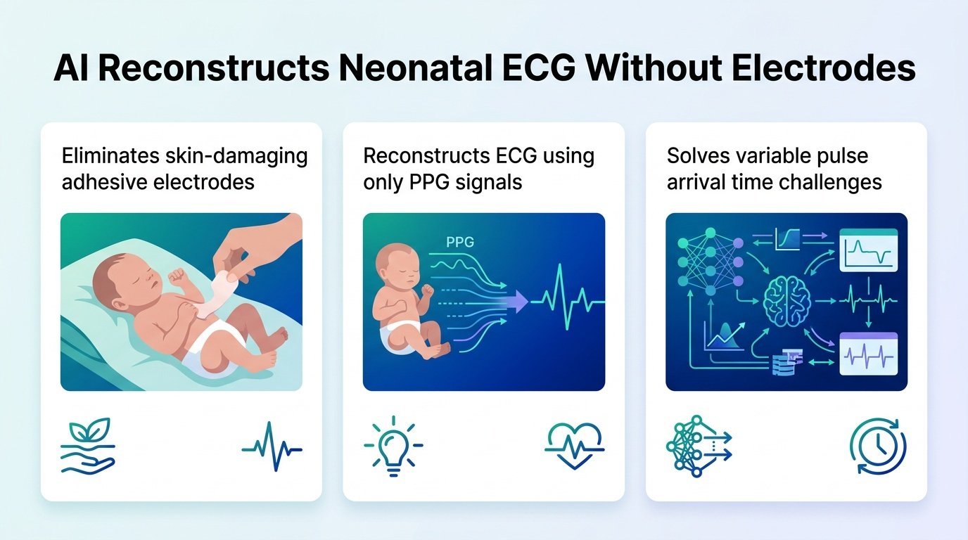
A new AI model reconstructs missing neonatal heart signals using light-based sensors, bypassing the need for irritating skin adhesives in intensive care.
How do you monitor a premature baby’s heart when the very stickers used to track it can tear their paper-thin skin? This is the daily dilemma in neonatal intensive care units. Traditional electrodes cause painful skin injuries, but shifting to light-based sensors has historically failed because babies have highly unpredictable blood flow.
This disconnect is where standard algorithms fall short. By abandoning the obsession with perfect signal alignment, this new model proves that AI can handle the chaotic biology of a newborn. It challenges the assumption that medical AI needs perfectly synchronized inputs to be clinically useful.
Why babies confuse standard AI
Adult algorithms assume a predictable delay between a heartbeat and the pulse wave reaching the skin. In newborns, this pulse arrival time shifts constantly. Trying to force these signals to align artificially ruins the diagnostic data. The researchers bypassed this by designing an alignment-free model that learns the natural, fluid relationship between blood flow and electrical heart activity.
To train the system, researchers used 52,566 ten-second windows of concurrent signal data. This dataset came from 159 neonatal intensive care patients. Crucially, they split the data at the patient level. This means the AI was tested on entirely new babies, proving it learned universal physiological patterns rather than just memorizing individual heart signatures.
Reconstructing the missing beats
Instead of forcing synchronization, the model integrates light-based hemodynamic timing with lead-specific heart context. The AI filled in missing electrical heart signals with high precision, even during simulated sensor dropouts. This resilience is vital because physical movement constantly disrupts sensors in real incubators.
The model demonstrated high accuracy across several simulated data loss scenarios:
- Under a 40% random missing data condition, it achieved a Pearson correlation coefficient of 0.96, a mean absolute error of 0.04, and a root mean square error of 0.07.
- During a 4.0-second continuous block loss—a massive gap for a newborn’s rapid heart rate—it maintained a correlation of at least 0.90.
- Under a severe 60% random patch loss, it still held a correlation of at least 0.90.
The path to clinical use
This approach could allow hospitals to maintain continuous heart monitoring even when physical sensors temporarily lose contact. If the electrical signal drops out, the light-based sensor steps in to fill the gap. However, we must be realistic about the current stage of this technology. This was a retrospective study on historical data, not a live trial in a chaotic ward.
Before doctors can make real-time diagnostic decisions based on reconstructed signals, the model requires prospective validation in active intensive care units. Until then, it remains a highly promising proof of concept for gentler pediatric care.
Read the full study in medRxiv.