
A zero calcium score often gives patients false reassurance, but a new AI model uncovers deadly vascular inflammation hidden inside standard noncontrast CT scans.
A clear Coronary Artery Calcium (CAC) scan often sends patients home without treatment. That clinical habit leaves a major blind spot, as significant numbers of cardiac events occur in people deemed low risk by calcification alone.
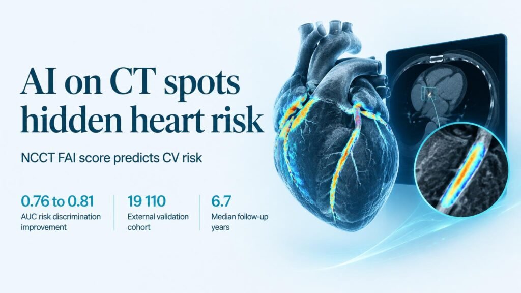
Building on landmark findings from the CRISP CT study, researchers evaluated an artificial intelligence algorithm that measures the Fat Attenuation Index (FAI) directly from routine noncontrast CT scans. The software extracts inflammatory signals from pericoronary fat without requiring contrast dye or additional imaging protocols.
Testing the AI model
The researchers trained the AI model in a derivation cohort of 6,818 individuals with a median age of 61 years (52.0% female). They validated its performance across an independent clinical cohort of 19,110 patients with a median age of 56.0 years (48.6% female). Over a median follow-up of 6.7 years between 2010 and 2024, the dataset recorded 536 cardiovascular deaths and 1,727 major adverse cardiovascular events.
Adding the noncontrast FAI score to standard calcium scoring improved risk discrimination, raising the area under the curve from 0.76 to 0.80 (P < .001). Contrast CT FAI achieved 0.81, proving noncontrast scans can deliver nearly identical prognostic accuracy.
The risk reclassification was most stark among patients with low calcification. Among cardiovascular deaths in patients with a CAC score of 0, 44.9% had a noncontrast FAI score above the 75th percentile. Similarly, 44.2% of deaths in the low CAC group (scores 1 to 99) had high FAI scores.
Rethinking low-risk labels
Inflammation drastically altered the real-world event rates of seemingly safe patients.
For patients with zero CAC, an elevated noncontrast FAI score above the 75th percentile pushed cardiovascular mortality from 1.4 to 4.0 per 1,000 person-years. Major adverse cardiovascular events in this same group rose from 5.9 to 13.3 per 1,000 person-years.
These findings mirror outcome data from the ORFAN multicentre cohort study, confirming that vascular inflammation drives heart attacks independent of plaque buildup. Evaluating noncontrast scans allows clinicians to flag at-risk individuals during routine screening.
What needs to happen next
Risk identification is not proof of treatment efficacy. Observational cohorts show that noncontrast FAI improves risk reclassification, but they do not prove that starting therapy solely due to a high FAI score reduces deaths.
Randomized clinical trials must now determine whether initiating targeted anti-inflammatory or aggressive lipid-lowering therapy in high-FAI, zero-CAC patients changes clinical outcomes before health systems adjust standard prescribing guidelines.
Read the full paper in JAMA Cardiology.