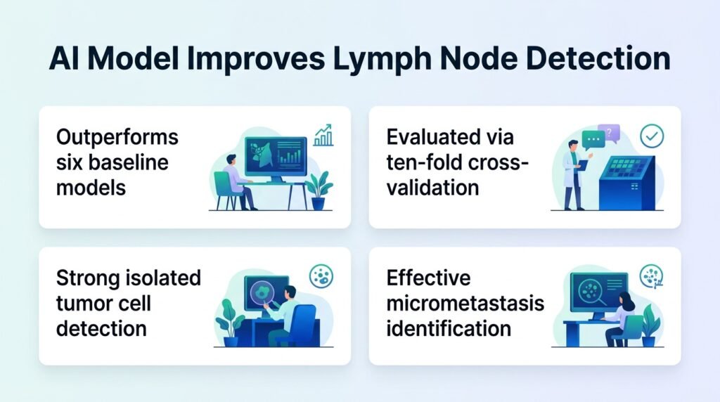
A new artificial intelligence model outperforms existing tools in spotting tiny cancer deposits in lymph nodes across multiple cancer types.
Pathologists routinely miss the microscopic clusters of cancer cells that signal a tumor has begun to spread. These tiny deposits, known as micro-metastases or isolated tumor cells, are incredibly easy to overlook on a massive digital slide. Missing them can lead to undertreatment and early relapse for the patient.
A new study published in Nature Communications introduces a model called MambaMIL+HiLA-MIL to solve this diagnostic blind spot. The research challenges the prevailing assumption that pathology AI must be highly specialized for single organs. Instead, this single model successfully classifies lymph nodes across multiple cancer types without sacrificing accuracy.
The analytical shift
For years, computational pathology has relied on narrow models trained on single organs. Early work in Nature Biomedical Engineering proved that weakly supervised learning could analyze whole-slide images without tedious manual annotations. Later, researchers successfully targeted specific organs, such as colorectal cancer in Modern Pathology. This new model consolidates those efforts into a single, multi-cancer tool.
This consolidation is critical. Running separate AI models for every organ type in a busy clinical lab is a logistical nightmare. A unified model simplifies the workflow and lowers the computational barrier to entry for hospitals. It shifts the focus from narrow, bespoke algorithms to unified clinical diagnostic pipelines.
How the model performed
The researchers combined Vision Mamba with a high-low attention separation mechanism. They evaluated the model using a rigorous 10-fold cross-validation against six baseline models. The testing spanned four different feature extractors: ResNet, UNI, Virchow, and GigaPath.
- The model significantly outperformed all six baseline systems.
- It accurately classified slides into four distinct categories, ranging from negative cases to isolated tumor cells and macro-metastasis.
- Advanced foundation models, specifically UNI and GigaPath, yielded significantly better performance than older extractors like ResNet and Virchow.
- The system maintained high stability across multiple clinical centers and diverse cancer types.
Why this matters
This finding is not just about beating baseline algorithms. It proves that modern foundation models like GigaPath can extract features rich enough to detect isolated tumor cells, which are often just a few pixels wide on a whole-slide image. The model maintains high performance on negative cases, meaning it will not overwhelm pathologists with false positives.
However, limitations remain. The study is retrospective and relies on existing datasets. We still do not know how the model handles the real-world variations in slide preparation and staining that occur daily in smaller, regional laboratories. Prospective clinical trials are needed to prove it actually reduces diagnostic error rates in real-time practice.
Read the full study in Nature Communications.