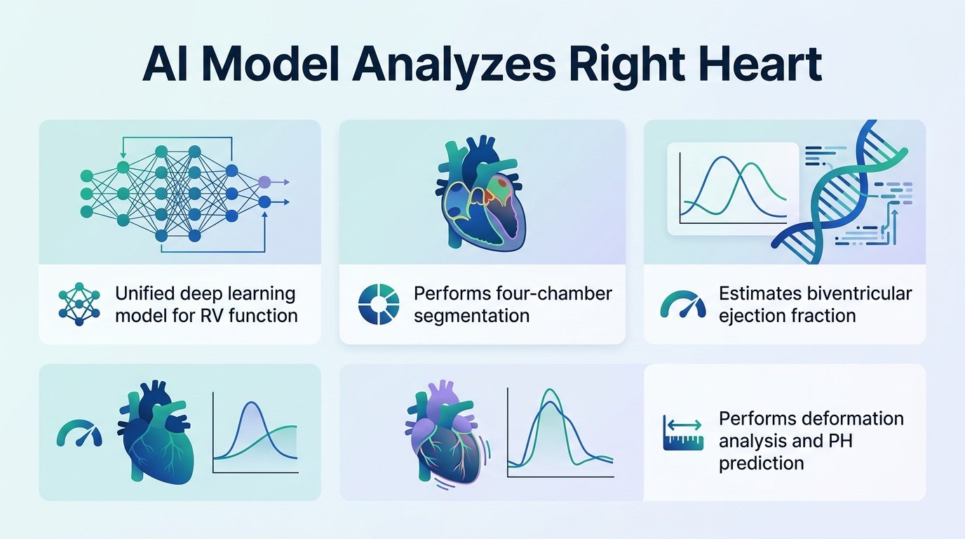
By consolidating complex cardiac measurements into a single view, a new deep-learning model challenges the clinical habit of ignoring the right side of the heart.
Why does cardiology treat the right side of the heart like a second-class citizen? For decades, left-ventricle metrics have dominated clinical software. The complex, crescent-shaped right ventricle has been dismissed as too difficult to model from basic ultrasound views, leaving patients with pulmonary hypertension in a diagnostic blind spot.
A new preprint introduces PH-ECHO-AI, a unified model designed to dismantle this imbalance. By extracting four-chamber segmentation, biventricular ejection fraction, and pulmonary hypertension predictions from a single apical four-chamber clip, it proves we do not need expensive 3D imaging to get a clear picture of right-heart strain. This challenges the assumption that right-ventricle tracking requires specialized, high-cost protocols. It aligns with recent efforts to simplify right ventricular assessment, such as the deep learning tools discussed in Echocardiography.
Researchers built the model using **8,416 clips** from four public datasets: EchoNet-Dynamic, CAMUS, RVENet, and MIMIC-IV-ECHO. The team evaluated the tool using held-out data. This included **1,416 clips** for segmentation, **600 clips** for function, and **1,076 MIMIC-IV patients** for pulmonary hypertension prediction. Of the function clips, **350** were referenced to 3D-echocardiographic right ventricular ejection fraction and **250** to the EchoNet left ventricular ejection fraction.
The power of one view
The results show that a single view can yield highly accurate structural data. The model bypassed traditional geometric assumptions to estimate right ventricular ejection fraction directly.
- Pooled Dice similarity coefficients reached **0.925** for the left ventricle, **0.836** for the right ventricle, **0.910** for the left atrium, and **0.904** for the right atrium.
- Left ventricular ejection fraction was estimated with a correlation of **r=0.845** and a mean absolute error of **4.67%**.
- Right ventricular ejection fraction reached a correlation of **r=0.754** and a mean absolute error of **4.98%**, vastly outperforming traditional geometric fractional area change which scored just **r=0.278**.
- Pulmonary hypertension was detected using geometry alone with an area under the receiver operating characteristic curve of **0.697** and a Brier score of **0.061**.
Real-world limitations
These numbers are promising, but the clinical reality demands caution. The right ventricular ejection fraction was evaluated on same-source, clip-disjoint data. This means true cross-centre validation for this metric is still missing. If the model cannot replicate these results on entirely foreign hospital scanners, its clinical utility drops.
Furthermore, an AUC of **0.697** for pulmonary hypertension prediction is modest. It is a useful screening tool, not a diagnostic replacement. However, the true value here is the consolidation of metrics like TAPSE, MAPSE, and strain into one automated step. This shifts the bottleneck from manual, highly variable expert tracing to instant, standardized triage.
Read the full study on medRxiv.