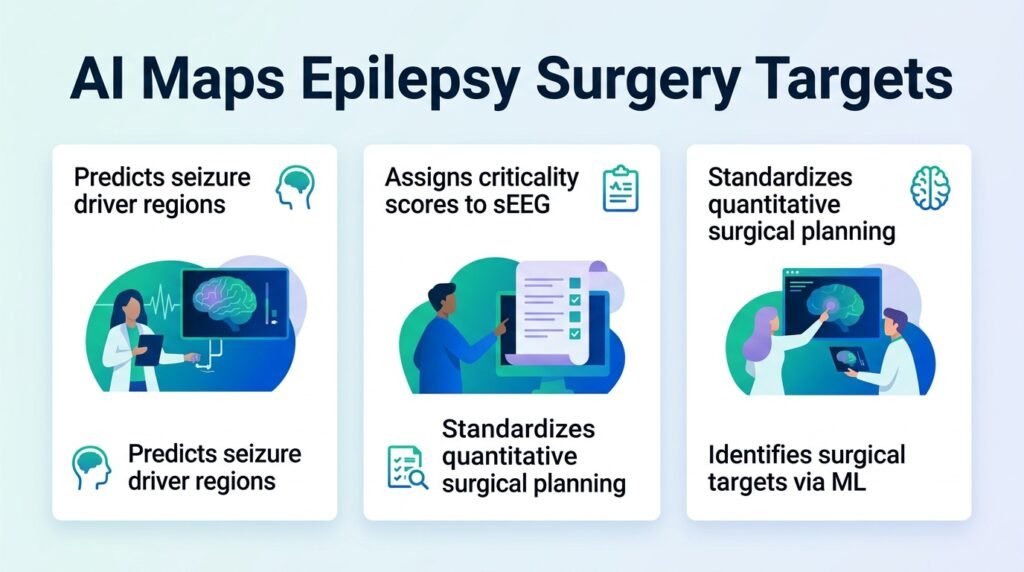
A new computational platform pinpointed compact brain targets to help surgeons stop drug-resistant seizures.
Epilepsy surgery is often a high-stakes guessing game. Surgeons insert depth electrodes into the brain, record days of electrical chatter, and then must decide exactly which tissue to cut out. If they miss the true driver of the seizures, the patient continues to suffer.
A new multicenter validation study of an algorithm called CN Suite suggests we can stop guessing. By mapping causal networks in brain signals, the software identifies highly specific, compact targets. This shifts the surgical paradigm from wide tissue resections to highly targeted, minimally invasive ablations.
Why compact targets matter
Researchers tested the algorithm on clinical data from 60 patients aged two and older across four U.S. Level 4 epilepsy centers. The algorithm had already been trained on an independent cohort of 37 patients and locked. The main goal was to see if the algorithm’s “criticality scores” matched the tissue removed in patients who actually got better.
The results showed a clear signal. Patients with favorable outcomes had significantly higher criticality values in their surgically treated tissue compared to those with poor outcomes, with an effect size of 0.74. The algorithm also showed that high-criticality contacts clustered tightly together. The nearest-neighbor distance was just 9 mm, compared to the 17 mm expected by chance.
This spatial tightness is the real story.
It means surgeons do not need to remove large swaths of brain tissue. Instead, they can use precise tools to destroy tiny, critical nodes. In fact, the software’s sensitivity was highest at 80% in small procedures involving 10 or fewer treated contacts. It performs best when the surgical footprint is kept small.
Explaining surgical failures
When epilepsy surgeries fail, doctors often wonder if they targeted the wrong region entirely. This analysis suggests a different answer. In patients whose surgeries failed, the high-criticality tissue identified by the AI remained just outside the surgical boundary.
This means the localization was likely correct, but the treatment coverage was incomplete. This finding gives clinicians a clear, actionable roadmap for reoperation rather than leaving them in the dark.
The limits of the math
No algorithm is perfect, and this tool has clear boundaries. Prediction specificity at the contact level was 84%. For the 28 cases analyzed for seizure-free contacts, 88% of identified contacts were indeed seizure-free.
Here are the key metrics from the validation trial:
- An effect size of 0.74 distinguishing favorable and unfavorable outcomes.
- A high contact-level specificity of 84%.
- An 80% sensitivity rate for small focal procedures.
- A tight 9 mm clustering of high-criticality targets.
However, the algorithm’s sensitivity decreased as resection size grew, meaning it is less useful for large, complex surgeries. The pediatric sample size was also small, meaning more data is needed before pediatric neurosurgeons can rely on it completely. Clinicians should view this as a tool for precision ablation, not a cure-all for diffuse brain disease.
Read the full study in medRxiv.