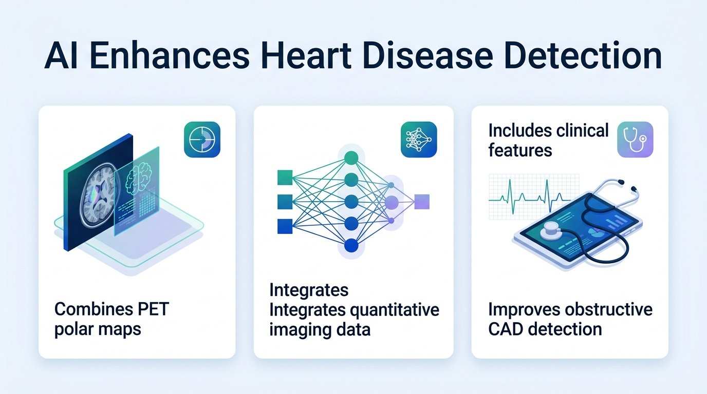
A new multimodal AI model outperforms standard PET scan metrics to catch blocked coronary arteries before they cause a heart attack.
How much critical data do cardiologists leave on the table when analyzing a heart scan? Positron emission tomography (PET) provides a wealth of data on blood flow and heart function. Yet clinicians often struggle to synthesize these complex imaging maps with a patient’s clinical risk factors in real time.
This diagnostic gap is where errors happen. By failing to connect the dots between visual perfusion maps and tabular clinical data, traditional analysis misses subtle signs of severe blockages. This study challenges the status quo by proving that a machine learning model can integrate these distinct data silos to outperform human reading and standard quantitative metrics.
This shift aligns with a broader movement toward integrated diagnostics. As explored in research on Changing Paradigms in the Diagnosis of Ischemic Heart Disease by Multimodality Imaging, combining anatomical and functional data is becoming essential for accurate cardiac care.
The data behind training
Researchers built a two-stage contrastive learning framework using the massive REFINE PET registry. In the first stage, they pretrained the model on 12,225 PET studies from eight sites to learn the relationships between 15-channel polar maps, blood flow measures, and clinical variables. The second stage fine-tuned the model on 968 patients who had confirmed invasive angiography results.
In this training cohort, 60% of patients had obstructive coronary artery disease, 66% were male, and the median age was 70. To prove the model works in the real world, researchers then tested it on an external validation cohort of 1,865 patients from six independent sites. In this validation group, 55% had obstructive disease, 64% were male, and the median age was 67.
Key diagnostic performance gains
- The AI model achieved an area under the curve (AUC) of 0.85 for detecting obstructive coronary artery disease.
- At a matched specificity, the AI achieved 89% sensitivity compared to only 85% for conventional visual stress scores.
- The negative predictive value rose to 81%, up from 73% in standard clinical evaluations.
- The model achieved an overall net reclassification improvement of 8.9%.
The real-world clinical payoff
These numbers are not just minor statistical bumps. Raising the negative predictive value to 81% means doctors can confidently rule out major blockages without sending patients for invasive, risky angiograms. This addresses a long-standing challenge in cardiac imaging, where precise quantification across different clinics remains difficult, as documented in studies of Multimodality Quantitative Assessments of Myocardial Perfusion.
However, the study has limitations. The data is retrospective, and all patients in the validation group had already been referred for invasive angiography. This creates a selection bias, meaning the AI must still be tested in a broader, unselected screening population before widespread clinical adoption.
Read the full study in medRxiv.