 Yusuf Yesil, MD, MIS
Sarcopenia significantly worsens overall survival in gastric cancer patients. Yet physical screening requires grip strength and gait speed tests that busy surgical clinics rarely perform. That friction leaves high-risk patients unidentified until post-operative complications hit.
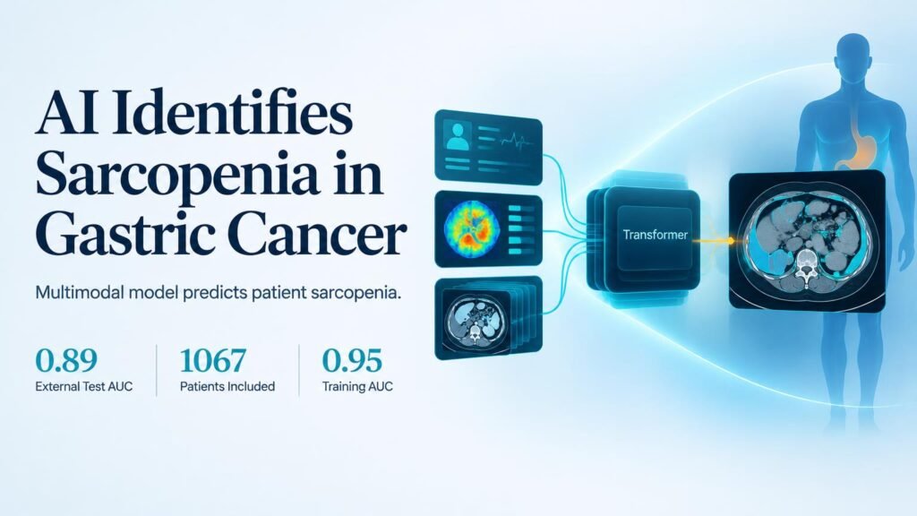
A new trial bypasses physical testing entirely. Researchers built a multimodal transformer model that combines clinical data, radiomics, and 2.5D deep learning features from standard abdominal CT scans. By automating screening directly from routine pre-operative imaging, the software turns existing diagnostic workflows into prognostic tools.
That shifts sarcopenia detection from an active physical exam to a passive imaging analysis.
Multimodal Fusion Beats Single Modalities
The study evaluated 1067 patients with gastric cancer across two medical centers. The cohort included 782 male (73.3%) and 285 female (26.7%) patients, with consistent gender distribution across training, validation, and external test cohorts (p = 0.34). Manual diagnosis required confirming low skeletal muscle index at the L3 vertebral level alongside low grip strength or low gait speed for severe cases.
Single-modality tools showed obvious performance ceilings. Combining these features inside an integrated architecture yielded much stronger discriminative power across validation and external cohorts.
- The L3 radiomics model achieved AUCs of 0.85 in validation and 0.80 in external testing.
- A 2.5D ResNet50 deep learning model reached AUCs of 0.81 in validation and 0.83 in external testing, outperforming its 2D counterpart.
- An XGBoost fusion baseline recorded AUCs of 0.85 in validation and 0.86 in external testing.
- The final transformer sarcopenia model reached AUCs of 0.95 in training, 0.87 in validation, and 0.89 in external testing for sarcopenia, alongside 0.84 for severe sarcopenia in external testing.
What the Performance Gains Mean
This work builds on previous efforts to link tissue composition with patient outcomes. Earlier studies showed that CT radiomics of skeletal muscle can detect sarcopenia in upper gastrointestinal tumors. Subsequent research demonstrated that radiomic feature extraction predicts prognosis in gastric cancer cohorts. This new transformer model demonstrates that combining clinical metrics with 2.5D spatial features improves stability across external hospital settings.
Yet high area-under-the-curve metrics do not equal automated clinical replacement. Retrospective validation across two centers confirms retrospective accuracy, not real-world clinical intervention.
The core limitation is operational. While an AUC of 0.89 proves the software identifies muscle degradation on static scans, it cannot measure functional physical reserve or dynamic recovery. Care teams must still prove that acting on these automated AI risk scores through nutritional or physical interventions actually improves overall survival.
Surgical teams should view this tool as an automated triage trigger on pre-op imaging, not a standalone diagnostic replacement.
Read the full paper in the Journal of Cachexia, Sarcopenia and Muscle.