A new deep learning model proves that artificial intelligence does not just mimic old diagnostic rules to spot deadly heart valve disease—it finds hidden signals doctors have been missing for decades.
For years, skeptics argued that AI models reading electrocardiograms were just doing basic math in disguise. Critics assumed these algorithms simply calculated left ventricular hypertrophy (LVH)—the thickened heart muscle caused by overwork—and rebranded it as valve disease detection. If true, AI would be a redundant, expensive mirror of existing clinical rules.
This new study dismantles that assumption.
By proving that AI bypasses traditional LVH criteria, the research establishes AI-ECG as an independent diagnostic tool. It challenges the medical community to rethink what an ECG can actually reveal about structural heart disease.
The data behind the model
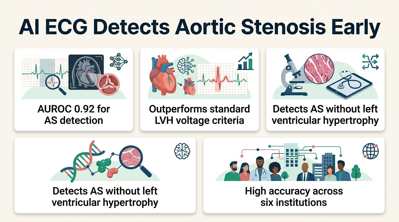
Researchers built and tested the deep learning model using a massive dataset of 244,816 ECGs from 51,713 patients. The data came from the CLIDAS database, which spans six academic institutions in Japan. To train the algorithm, the team matched these ECGs with inpatient Diagnosis Procedure Combination codes to identify cases of aortic stenosis.
The model outperformed traditional diagnostic metrics by a wide margin. The performance gap shows that human-designed rules are leaving critical data on the table.
- The AI model achieved an area under the receiver operating characteristic curve (AUC) of 0.849, with a tight confidence interval of 0.832-0.865.
- At a threshold of 0.1, the model demonstrated 79.1% sensitivity, 73.9% specificity, and a 98.0% negative predictive value.
- Traditional LVH voltage criteria lagged far behind, with the Sokolow-Lyon index scoring an AUC of 0.706 and the Cornell index scoring 0.692.
- Adding traditional LVH criteria to the AI model provided no incremental benefit, resulting in an AUC of 0.849 versus 0.847.
Why this finding matters
If the AI merely tracked muscle thickness, it would fail to catch early-stage aortic stenosis patients whose heart walls have not yet thickened. Instead, explainable AI imaging (Grad-CAM) showed the model focused on QRS complexes in the limb leads. This is a distinct electrical region from the chest leads typically used to calculate LVH.
This means the AI is identifying unique electrical signatures of valve obstruction, not just the muscle damage that follows it. This capability aligns with other recent advances, such as using an ensemble deep learning algorithm for structural heart disease screening to catch silent cardiac issues.
With a negative predictive value of 98.0%, this tool is a highly reliable gatekeeper. Clinicians can confidently use it to rule out aortic stenosis, saving patients from unnecessary and costly echocardiograms. It builds on previous research demonstrating how neural networks classify complex cardiac phenotypes, including studies on hypertrophic heart diseases using electrocardiograms.
The clinical reality check
Despite the strong performance, some limitations remain. The model relied on administrative diagnostic codes rather than direct echocardiographic measurements for its ground truth. Additionally, as a retrospective study, it still requires prospective validation in diverse, real-world clinical workflows before widespread adoption.
Read the full preprint study on medRxiv.