Automating postoperative wound checks could save clinic hours, but a new trial reveals the steep performance drop-off when algorithms meet real-world patients.
How do you monitor a surgical incision once the patient is already home? Fast-track recovery protocols mean patients leave the hospital earlier than ever, shifting the burden of spotting infections to at-home telemedicine. But manual review of wound photos is slow, inconsistent, and prone to human error.
This is where computer vision steps in, promising to filter out normal healing from active infections. Yet, the transition from pristine training data to messy clinical reality is rarely smooth.
This study highlights a classic machine learning bottleneck: the generalization gap. While the model excelled on its internal test data, its accuracy dipped when tested on a completely new patient population. This drop-off challenges the assumption that a model trained on historical databases is immediately ready for diverse, real-world clinics.
For clinical leaders, this means AI cannot yet act as a solo diagnostic tool. Instead, its immediate value lies in automated triage—filtering out obviously healthy wounds so human clinicians can focus on borderline cases. This aligns with broader efforts to integrate smart monitoring tools into post-surgical recovery, as discussed in recent work on vascular health care technology.
Testing the algorithm
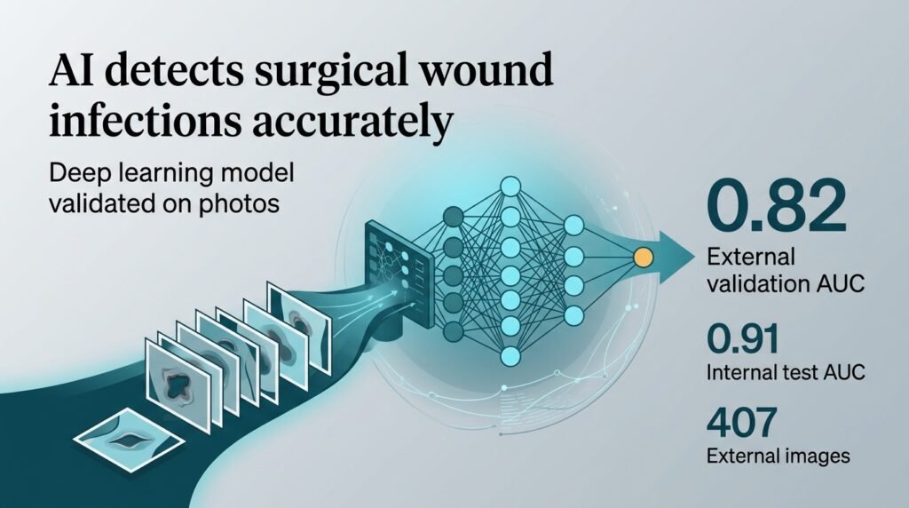
Researchers built the deep learning model using an InceptionV3 convolutional neural network architecture. They trained the system on a retrospective dataset of 4,978 wound images, split into training (70%), validation (15%), and internal test (15%) sets. To test how the model performs in the wild, they collected a prospective external validation set of 407 images from 95 patients at a large academic center.
The performance gap between the two environments was notable.
- In the internal validation set, the model achieved an area under the receiver operating characteristic curve of 0.91 (95% CI, 0.88-0.93).
- In the independent external validation set, the accuracy dropped to an AUC of 0.82 (95% CI, 0.75-0.90).
- Decision curve analyses still showed a positive net benefit for clinical utility.
The reality of triage
A drop from 0.91 to 0.82 AUC is the difference between an autonomous diagnostic tool and a supervised triage assistant. The model used gradient-weighted class activation mapping heat maps to show clinicians exactly where it was looking on the wound. This transparency is helpful, but the drop in external accuracy shows the algorithm still struggles with variations in lighting, camera angles, and skin tones found in different hospital systems.
Relying on patients to take high-quality photos at home introduces massive variability. If a model’s accuracy degrades under real-world conditions, hospitals risk either missing critical infections or overwhelming clinics with false alarms. To make telemedicine work, we must standardize how wound photos are captured before we can trust the algorithms that read them.
Read the full study in JAMA Surgery.