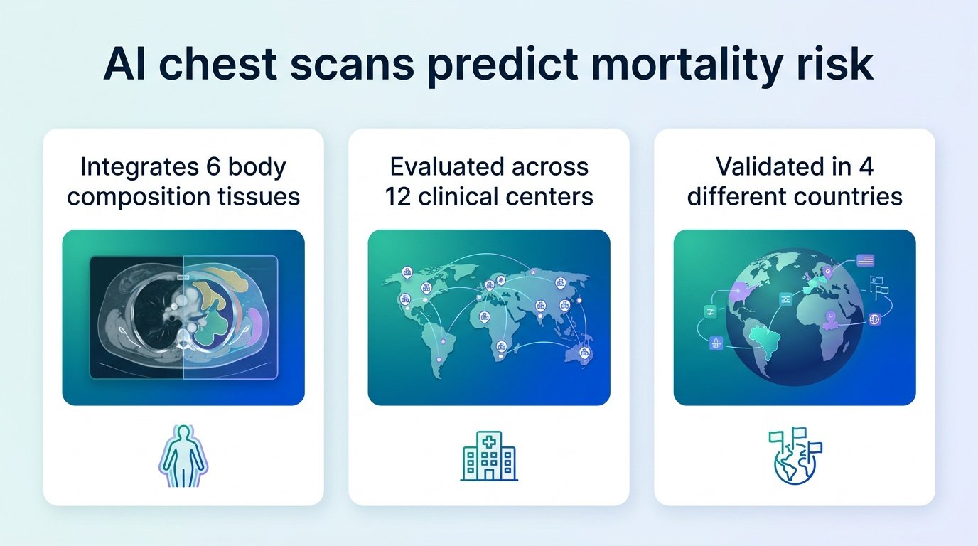
A new artificial intelligence index uses routine heart scans to calculate mortality risk, proving that how we store fat and muscle matters far more than simple body weight.
Clinicians still rely on Body Mass Index (BMI) to judge metabolic health, even though we know it is a blunt, deeply flawed instrument. It cannot tell the difference between heavy muscle and dangerous visceral fat. A new study challenges this status quo by turning routine, low-dose chest CT scans into a multi-dimensional map of survival.
This is not just about automated imaging. It is a shift in how we define patient risk. By extracting hidden data from scans already being performed, clinicians can spot high-risk patients without ordering expensive new tests. This moves opportunistic screening from a conceptual novelty into a practical tool for preventative care.
Researchers built and validated the Body Composition Index (BCI) using a dataset of 28,509 consecutive patients across 12 centers in four countries. All patients underwent myocardial perfusion imaging with routine low-dose chest CT attenuation correction scans. The team trained the AI model on 15,037 patients, integrating bone, skeletal muscle, four fat compartments, coronary calcium scores, and basic demographics. They then tested it on an internal cohort of 6,444 patients and an external cohort of 7,028 patients.
During a median follow-up of 3.5 years (with an interquartile range of 1.9 to 5.1 years), 4,697 patients died, representing 16% of the total cohort. In the external test group, the BCI predicted all-cause mortality with an area under the receiver operating characteristic curve of 0.78 (95% CI [0.76, 0.79]) and a Harrell concordance index of 0.75 (95% CI [0.73, 0.76]).
What the AI found
- Visceral adipose tissue attenuation was the most powerful predictor of death.
- Skeletal muscle volume was the second most influential marker.
- The index maintained its predictive power across different imaging protocols and patient subgroups.
Why this matters
This finding forces us to rethink standard diagnostic pathways. We are already collecting this imaging data during routine cardiac workups. Leaving it unanalyzed is a missed medical opportunity. This approach builds on previous work showing the value of opportunistic imaging, such as using AI body composition in lung cancer screening to look beyond oncology.
However, the real clinical hurdle is integration. Doctors do not need another isolated score to copy into an electronic health record. For this index to succeed where others failed, it must be embedded directly into automated radiology workflows, much like the volumetric models discussed in The Lancet Digital Health.
We must remain cautious about the study’s limitations. This was a retrospective analysis of patients already undergoing cardiac imaging, meaning they likely had higher baseline cardiovascular risks than the general public. Additionally, while the model simulated how improving body composition could reduce mortality, real-world clinical trials are still needed to prove that changing these tissue metrics actually extends lives.
Read the full study in medRxiv.