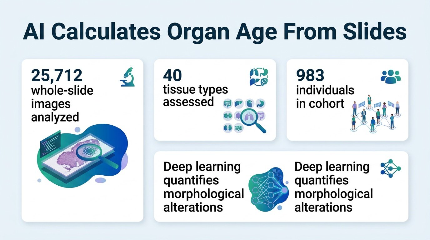
By training deep learning models to read structural decay in tissue biopsies, researchers can now estimate the biological age of specific organs and predict chronic disease risk.
How old are your lungs compared to your liver? We usually measure aging through systemic markers like blood pressure or gray hair, but our organs decay at wildly different rates. This invisible, uneven decline is why some people suffer sudden strokes at fifty while others maintain sharp cognitive health well into their eighties.
A new study challenges the assumption that aging is a uniform, body-wide clock. By analyzing microscopic tissue architecture, researchers have proved that structural wear-and-tear is a highly localized process. This shifts the focus of longevity medicine from systemic treatments to targeted, organ-specific interventions. It forces us to rethink chronological age entirely, suggesting that personalized medicine must address the body as a mosaic of different biological ages.
To build these localized clocks, researchers analyzed a massive dataset of 25,712 whole-slide histopathological images. These samples spanned 40 different tissue types collected from 983 individuals in the Genotype-Tissue Expression cohort. Using deep learning, the team quantified nuanced morphological alterations to develop “tissue clocks” that measure structural integrity. These digital clocks successfully mapped physical decline to known biological aging markers, including telomere attrition and subclinical pathologies.
Reading organ age in blood
- The AI successfully predicted biological age across 40 distinct tissue types based on structural changes.
- These tissue-specific clocks correlated directly with physical markers of decline, such as telomere attrition and comorbidities.
- By pairing histology with transcriptomic data, the models predicted organ-specific age gaps directly from blood samples.
- The researchers validated these aging signatures across independent cohorts for eight prevalent diseases, including Alzheimer’s disease, stroke, and Crohn’s disease.
The real value of this research lies in the bridge built between deep tissue structure and blood chemistry. By pairing histology with transcriptomic data, the researchers created a method to predict organ-specific age gaps from a standard blood draw. This means doctors could eventually screen for silent organ decay without invasive biopsies. If a blood test can flag an aging brain years before cognitive decline begins, the window for preventative therapy opens dramatically.
The limits of tissue clocks
However, clinical adoption faces immediate hurdles. The initial model relies heavily on the Genotype-Tissue Expression cohort, which may not represent global genetic and lifestyle diversity. Furthermore, translating these complex structural patterns into routine clinical decisions requires standardized imaging pipelines that most hospitals currently lack. Until we can easily run these models on standard biopsies, the technology remains a powerful research tool rather than a diagnostic staple.
Read the full study in Nature Medicine.