 Yusuf Yesil, MD, MIS
Craniomaxillofacial surgery relies heavily on CT scans. That dependence keeps patients tied to specialized hospitals for every evaluation, exposing children to repeated radiation just to track surgical progress. Tele-Cranio shifts the initial assessment from hospital hardware to a standard mobile camera.
Early teleconsultation efforts in reconstructive surgery struggled because standard video calls cannot show underlying skeletal defects, a challenge highlighted in early research on perspectives of teleconsultation in craniomaxillofacial surgery. Tele-Cranio bridges that gap by mapping 2D facial soft tissue directly to underlying bone structures.
That shift changes how surgical teams approach remote triage.
Reconstructing Bone From 2D Photos
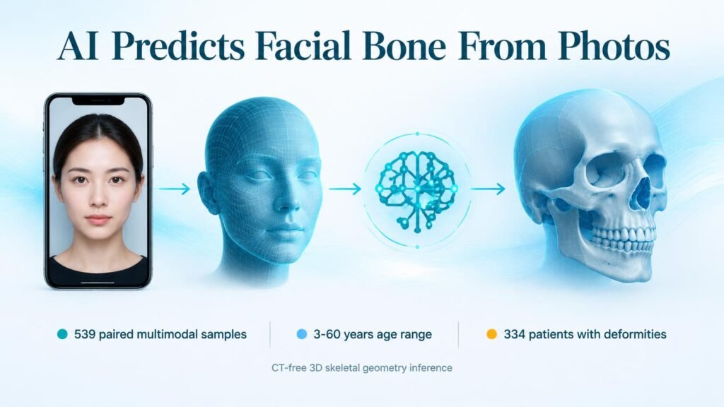
The research team built Tele-Cranio using 539 paired multimodal samples combining 2D facial photographs, 3D surface scans, and CT-derived skeletal models. The training cohort spanned individuals aged 3 to 60 years and included 334 patients with complex CMF deformities.
The dataset captured four major pathological groups: malocclusion, cleft lip with or without palate, hemifacial microsomia, and rare anatomical anomalies. Its underlying model learns a shared anatomical representation that links soft tissue directly to bone across both healthy and altered anatomy.
The system converts 2D photos into 3D surface meshes before estimating patient-specific 3D skeletal geometry. Once a surgeon enters a proposed bone modification plan, the tool generates an individualized 3D preview of the projected facial outcome.
Recent studies demonstrate that AI modeling can predict face shape based on underlying bone. Tele-Cranio flips this logic by inferring internal skeletal framework from external photos.
Screening Layer, Not CT Replacement
- Reconstructs 3D facial soft tissue and estimates underlying skeletal geometry from 2D photos.
- Evaluated across 334 deformity patients covering four distinct anatomical defect categories.
- Simulates postoperative facial appearance based on surgeon-defined skeletal modifications.
Tele-Cranio is a screening and communication layer rather than a replacement for diagnostic imaging. Retrospective estimates from surface photos cannot match the spatial resolution required for final operative execution.
The immediate operational value lies in pre-CT remote triage and radiation-free follow-up consultations. By letting surgical teams model outcomes and screen candidates remotely, health systems can reserve high-radiation CT scans for definitive surgical planning.
Read the full study in npj Digital Medicine.