 Yusuf Yesil, MD, MIS
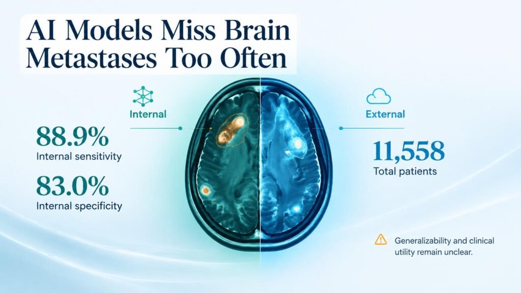
Distinguishing glioblastoma from brain metastasis on MRI scans dictates surgical planning and oncology treatment. Misclassifying these lesions leads to inappropriate surgical resection margins and delayed chemotherapy. While software developers claim quantitative imaging solves this visual overlap, pooled trial data reveals a significant gap between laboratory promises and clinical readiness.
A systematic review published in the Journal of Medical Internet Research analyzed 39 studies covering 11,558 patients to evaluate radiomics models. The findings show that while algorithms achieve reasonable accuracy inside initial trial settings, their performance degrades when exposed to unseen medical centers. This drop highlights how sensitive quantitative imaging features remain to scanner variability and local imaging protocols.
Performance Drops Beyond Training Labs
For standard binary classification between glioblastoma and metastasis, internal validation produced a pooled sensitivity of 88.9% and specificity of 83.0%. When tested on independent external validation cohorts, sensitivity fell to 83.8% and specificity dropped to 80.7%.
That drop in sensitivity marks the exact line where research metrics fail real-world patient safety.
The diagnostic risk sharpens in complex differential diagnoses. When models attempt multiclass differentiation involving primary central nervous system lymphoma, accuracy for secondary brain tumors collapses. Although prior work in multiclass brain tumor classification demonstrated early technical feasibility, aggregate evidence shows software fails to catch nearly three out of ten metastases in multi-disease differential scenarios.
Key Accuracy Metrics
- Binary external performance: Reached 83.8% sensitivity and 80.7% specificity across independent external test sites.
- Multiclass metastasis sensitivity: Fell to 70.6%, creating an unacceptably high rate of false negatives for metastatic disease.
- Lymphoma identification: Achieved a high specificity of 94.4%, but sensitivity reached only 75.0%.
- Multiclass glioblastoma accuracy: Showed a sensitivity of 84.4% alongside a specificity of 82.5%.
Why Clinical Utility Remains Unproven
These figures challenge assumptions that algorithmic feature extraction can replace invasive diagnostic pathways. While studies using automated machine learning demonstrate potential in tightly controlled cohorts, broad generalizability remains unverified. Critical methodological flaws evaluated via the Radiomics Quality Score 2.0 continue to limit the evidentiary value of published literature.
Retrospective diagnostic accuracy does not prove clinical efficacy or safer patient management. Retrospective single-center datasets inherently mask variations in slice thickness, magnet strength, and contrast timing across health systems.
Hospital leaders should resist deploying radiomics algorithms as autonomous diagnostic tools for intracranial masses. Until prospective multi-center trials validate these models in consecutive patient streams, radiomics outputs should remain strictly secondary exploratory indicators rather than drivers of surgical decisions.
Read the full meta-analysis in the Journal of Medical Internet Research.