 Yusuf Yesil, MD, MIS
Targeted lung cancer therapy depends on knowing a patient’s genetic status fast. Standard tissue biopsies take time and carry clinical risks, which leads many teams to test non-invasive radiomics. Yet standard imaging models often treat tumors as uniform spheres, missing critical biological variations inside and outside the lesion mass.
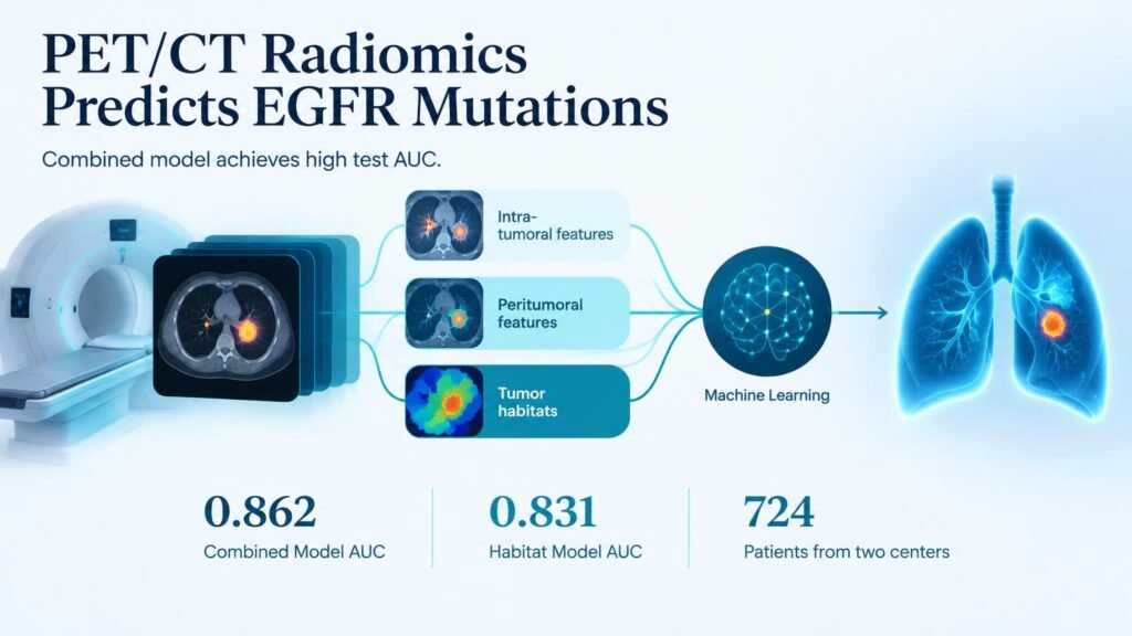
A two-center trial published in Frontiers in Medicine challenges that simplified approach. Researchers mapped micro-environments—or habitats—within lung adenocarcinoma tumors using baseline 18F-FDG PET/CT scans. By analyzing internal clusters and expanding tumor perimeters, the model captured subtle patterns that single-region algorithms routinely miss.
This work builds on prior attempts to decode tumor genetics from baseline imaging, such as multicentric radiomics models predicting EGFR mutations and early clinical-radiomic fusion techniques. The breakthrough here lies in mapping micro-habitat heterogeneity rather than averaging signal intensity across the entire lesion.
Predictive Accuracy and Spatial Highlights
The trial evaluated 724 patients across two independent hospital centers, dividing them into training, validation, and test cohorts. Investigators used software to extract features from internal tumor subregions (identified via k-means clustering) and peritumoral borders expanded by 2 mm to 8 mm radially.
- The combined model hit a top diagnostic performance with an AUC of 0.862 (95% CI: 0.80–0.93) in the independent test cohort.
- The stand-alone habitat model followed closely with an AUC of 0.831 (95% CI: 0.76–0.90), significantly outperforming standard single-region models (all p < 0.05).
- Among border-expansion models, the 6 mm radial extension yielded the highest predictive score around the tumor edge.
- SHAP explainability analysis showed that 16 out of 17 key features in the habitat model came from Habitat 1 and 2 subregions, while roughly two-thirds of the top predictive features were CT-based rather than PET-based.
Limits and Practical Reality
An AUC of 0.862 shows strong diagnostic correlation, but it is not a direct replacement for tissue sequencing. Retrospective validation across two centers proves technical feasibility, not improved patient survival or reduced time-to-treatment in actual workflow settings.
The operational takeaway is shift-focused: radiologists should stop relying exclusively on whole-tumor metabolic averages. Incorporating 6 mm peritumoral margins and CT structural subregions provides clear, interpretable signals that can triage high-risk patients for priority genetic testing when biopsy tissue is scarce.
Read the full paper in Frontiers in Medicine.