 Yusuf Yesil, MD, MIS
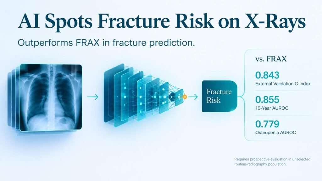
Standard screening tools miss a crucial window for intervention. The standard Fracture Risk Assessment Tool (FRAX) paired with bone mineral density (BMD) scans often fails to catch patients in the osteopenic range, where the majority of actual fractures occur. A deep learning survival model detailed in NEJM AI changes this dynamic by pulling predictive signals directly from routine chest radiographs, outperforming standard clinical calculators without adding patient scan time or radiation exposure.
This approach fundamentally challenges how health systems route patients for bone health evaluations. Instead of relying solely on primary care referrals for dual-energy X-ray absorptiometry (DXA), radiology workflows could flag high-risk individuals passively.
Stronger predictive power across cohorts
Researchers developed a convolutional neural network survival model trained on 53,109 adults at a single tertiary hospital. They then validated the algorithm externally across 85,284 patients from two independent institutions. The external cohort had a mean age of 58.6 years, was 68.9% female, and was followed for a median of 77 months. All participants had a chest radiograph taken within 1 year of a DXA scan.
The deep learning model consistently outperformed traditional risk scoring mechanisms across all time horizons:
- Achieved a 10-year censoring-adjusted AUROC of 0.855 compared to 0.785 for FRAX with BMD (P<0.01).
- Reached a overall C-index of 0.843 versus 0.779 for FRAX with BMD (P<0.01).
- Adding FRAX clinical data to the deep learning model yielded almost no additional gain, pushing the C-index only to 0.847.
- In the key osteopenia subgroup (35,358 patients), the model achieved a 10-year AUROC of 0.779 compared to just 0.659 for FRAX with BMD (P<0.01).
The performance gap in the osteopenic range is the key finding. By pulling subtle structural features from routine chest scans, the algorithm identifies vulnerable individuals that standard clinical metrics routinely classify as low or moderate risk.
Key limitations and clinical next steps
Superior discrimination on retrospective data does not automatically prove a software tool improves clinical outcomes or reduces fracture rates. A significant design constraint limits immediate deployment: every participant in this study was already referred for densitometry. This referral bias means the cohort does not match an unselected, general population walking into an outpatient clinic for a routine chest X-ray.
Before health systems use this tool for passive case-finding, prospective trials in broad, unselected cohorts are required to establish true positive predictive values and real-world clinical utility.
Read the full study at NEJM AI.