

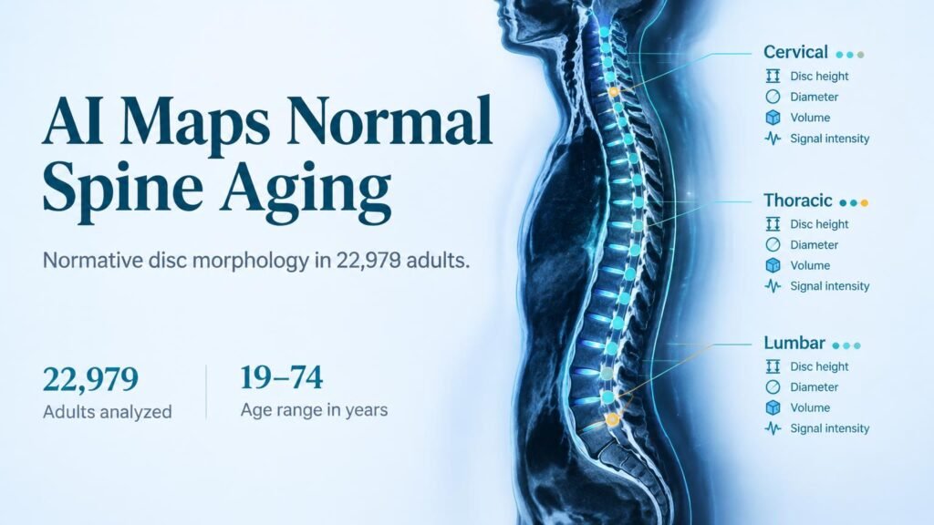
Automated MRI analysis of tens of thousands of asymptomatic spines challenges how radiographers distinguish normal structural aging from painful spinal pathology.
Radiologists often struggle to decide whether a flattened or widened spinal disc reflects disease or routine aging. Manual measurements across large populations are too slow to set clear baseline standards. By deploying an automated machine learning tool, researchers built a massive population reference for healthy spine morphology.
That baseline is the real value of this dataset.
This automation shifts AI from a simple diagnostic classifier to a baseline architect. Instead of guessing if a single scan shows early disease, automated pipelines can map normal physiological boundaries at scale. That reference framework is essential to stop clinicians from over-treating age-related disc changes that cause zero pain.
A massive automated imaging cohort
To establish these benchmarks, researchers analyzed T2-weighted MRI scans from **30,927** participants in the German National Cohort. After excluding individuals with back pain or osteoporosis, the final dataset included **22,979** healthy adults aged **19 to 74** years (**13,307** men and **9,672** women, with a mean age of **47.0 ± 12.5** years). An automated segmentation algorithm called SPINEPS measured intervertebral disc height, diameter, volume, and signal intensity across cervical, thoracic, and lumbar regions.
Earlier manual imaging research was constrained by small cohort sizes. Previous work includes an MR investigation of 169 cases evaluating thoracic disc shape and an analysis of 5,843 asymptomatic subjects focused solely on cervical signal intensity. Automating segmentation across all three spinal regions in over **20,000** subjects moves the field from small regional snapshots to whole-spine normative standards.
Key structural changes with age
The automated measurements revealed distinct structural trends across predefined age groups:
- Men consistently displayed higher disc height, diameter, and volume than women across all spinal segments.
- Increasing age strongly correlated with reduced disc height, volume, and signal intensity, alongside increased disc diameter.
- Higher body weight linked to increased signal intensity in the lower cervical spine, but showed an inverse relationship in the lower lumbar spine.
Limits and practical takeaway
The data demonstrate that disc expansion combined with volume loss is a predictable feature of aging. It should not automatically be classified as an active lesion. Radiologists must weigh these age-adjusted distributions before flagging structural wear as pathological.
However, the study design introduces an important selection bias. By excluding everyone with back pain, the cohort isolates asymptomatic aging but omits early structural changes that could predict future pain. The findings also rely on single-cohort data, so cross-scanner variability remains unverified.
For clinical teams, these population distributions provide a practical reference for surgical planning and diagnostic reports. Grounding scan interpretations in true population norms helps avoid unnecessary procedures for pain-free patients.
This research was published in European Radiology.