
Adding clinical data to ultrasound radiomics sharply improves breast cancer diagnostic accuracy, potentially sparing patients unnecessary biopsies.
Breast ultrasound often struggles with low specificity, leading to many invasive biopsies for lesions that turn out to be harmless. When radiologists classify a suspicious mass as BI-RADS 4a, the likelihood of malignancy ranges widely from 2% to 10%. That ambiguity drives high intervention rates and patient anxiety.
A new multi-center study shows that software relying on image features alone is not enough to fix this problem. Diagnostic precision shifts significantly only when algorithms pair quantitative imaging traits with routine patient metadata.
Combining clinical traits with radiomics
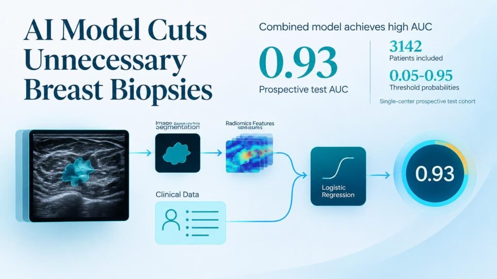
Researchers evaluated a cohort of 3,142 patients treated across eight hospitals between February 2012 and September 2024. They built five machine learning models using automatically extracted features from manually segmented ultrasound scans, followed by testing in an independent prospective single-center cohort.
The best standalone radiomics model, built on logistic regression, yielded modest area under the curve (AUC) performance: 0.83 in training, 0.82 in internal testing, 0.81 in external validation, and 0.82 in the prospective set. A model built strictly on patient clinical features actually outperformed radiomics alone, reaching AUCs of 0.87, 0.85, 0.87, and 0.86 across the respective cohorts.
The dynamic changed when researchers combined both streams. The combined clinical-radiomics tool achieved AUCs of 0.92 in training, 0.90 in internal validation, 0.92 in external validation, and 0.93 in prospective testing (p < 0.01 compared to single modalities). This mirrors broader efforts in the field, such as recent work evaluating ultrasound radiomic features for breast lesion differentiation.
Refining the BI-RADS 4a gray zone
The practical value of this approach centers on reclassifying borderline cases. Decision curve analysis showed a higher net clinical benefit for the combined model across threshold probabilities ranging from 0.05 to 0.95.
Crucially, applying the model to BI-RADS 4a cases increased the malignancy yield while maintaining high diagnostic sensitivity. This addresses a specific operational bottleneck by giving clinicians a reliable computational check before ordering invasive tissue sampling. Related research into machine learning for BI-RADS 4A lesions reinforces that targeted AI evaluation can reshape biopsy recommendations without missing active cancers.
Key Study Findings
- Enrolled 3,142 patients across eight institutions to train, validate, and prospectively test the diagnostic pipeline.
- Combined radiomics and clinical features achieved a prospective test AUC of 0.93, outperforming standalone radiomics (0.82) and clinical metrics (0.86).
- Using Shapley additive explanations (SHAP), the system provided interpretable feature scores to explain individual risk predictions to clinicians.
Limits to immediate adoption
These findings show clear diagnostic improvement, but operational barriers remain. The initial feature extraction required manual lesion segmentation, a time-consuming step that limits smooth integration into fast-paced radiology workflows.
Furthermore, while the development retrospective dataset was large and multi-centered, prospective validation occurred at only a single hospital. Health systems should view these results as a strong proof of decision support rather than justification to replace physician oversight or skip tissue confirmation today.
Study details published in Insights into Imaging.