
Machine learning on cerebellar MRI scans reveals that schizophrenia is not a single structural disease, explaining why standard drugs fail for many patients.
Treating schizophrenia as a uniform brain disorder leaves clinicians guessing why standard drugs help some patients while leaving others untreated. A broad machine learning analysis shows that cerebellar atrophy follows three entirely different biological paths. These distinct trajectories predict which patients resist standard drugs and who might benefit from non-invasive brain stimulation.
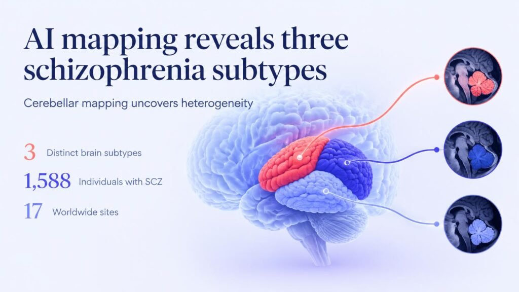
The cerebellum has long been ignored in psychiatric imaging compared to the cerebral cortex. Using a subtype and stage inference algorithm named SuStaIn, researchers analyzed cross-sectional 3D volumetric MRIs across 17 sites worldwide. The primary dataset matched 1,588 individuals with schizophrenia (638 females; mean age 31.9 ± 12.0 years) against 2,341 healthy controls (1,041 females; mean age 33.3 ± 13.6 years).
Three paths of brain decay
The model identified three spatiotemporal trajectories of cerebellar gray matter volume loss, which were successfully replicated in two independent cohorts of 1,334 and 530 patients. Each subtype stems from a unique origin point in the brain:
- Subtype 1: Originates in lobule X of the posterior lobe. It features gene enrichment in metabolic processes, severe emotion-related symptoms, and strong response to transcranial magnetic stimulation.
- Subtype 2: Originates in lobule III of the anterior lobe. It ties to immunity-related gene expression and shows poor response to antipsychotic medications.
- Subtype 3: Originates in lobule VIIb of the posterior lobe. It exhibits the most severe structural brain abnormalities, worse cognitive symptoms, and poor antipsychotic response.
That biological divide explains clinical failure points.
Standard antipsychotic trials often flatten these differences by treating all participants as a homogenous group. Longitudinal tracking of 381 patients over 12 months confirmed that subtypes 2 and 3 resist typical medication. Meanwhile, subtype 1 responds well to transcranial magnetic stimulation, offering a clear target for non-pharmacological care.
Limitations and clinical reality
These findings do not mean doctors can immediately order a brain scan to pick a schizophrenia prescription. The primary subtypes were inferred from cross-sectional brain images rather than lifetime longitudinal scans of individual progression. The treatment cohort of 381 patients remains small compared to the overall dataset, requiring prospective clinical trials before imaging can guide routine psychiatric dosing.
Subtyping shifts psychiatry from trial-and-error prescribing to targeted therapy. Mapping specific cerebellar sub-regions could soon stop clinicians from wasting months on ineffective drugs for non-responsive profiles.
Read the full preprint in medRxiv.