
A single facial photograph can now track biological aging and signal mortality risk in cancer patients.
Oncology clinics rely heavily on expensive genomic panels and complex scans to gauge patient prognosis. A new computer vision pipeline suggests a simple selfie might capture critical physiological decline just as well.
That visual signal is surprisingly hard to obscure.
By training on massive consumer image sets before fine-tuning on small clinical cohorts, researchers demonstrated that algorithmic facial analysis provides real prognostic value without requiring additional invasive blood draws or imaging.
Measuring age and survival
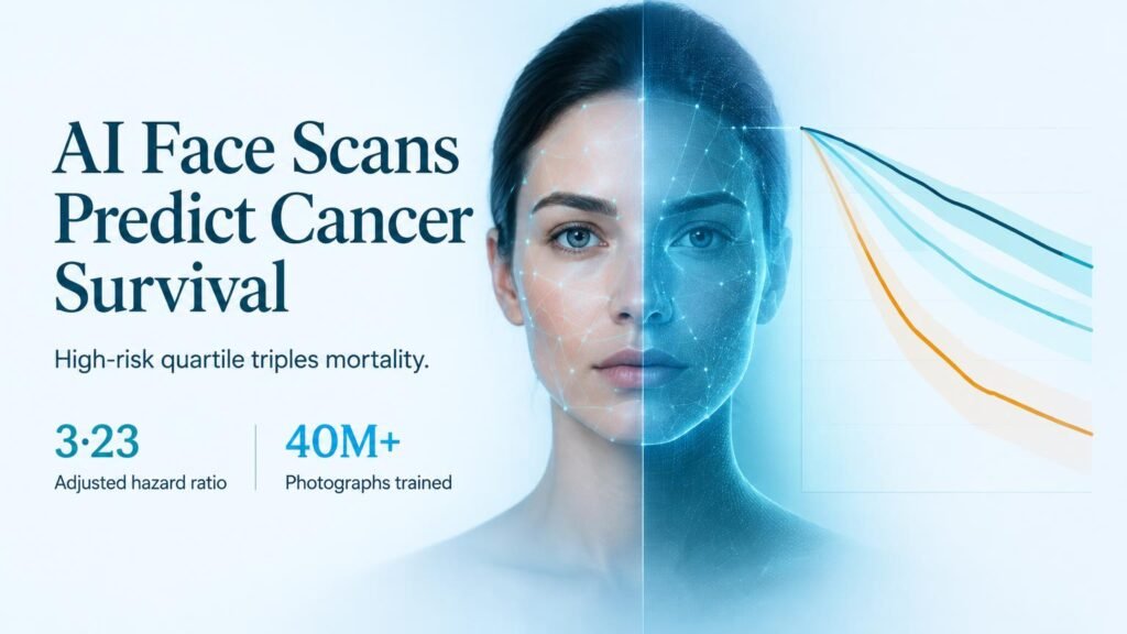
The foundation model, called FAHR-Face, was pre-trained on more than 40 million facial photographs before being split into two distinct tools. The first algorithm, FAHR-FaceAge, underwent two-stage fine-tuning across 10 public datasets to estimate biological age, achieving a mean absolute error of 5.1 years across the human lifespan.
The second algorithm, FAHR-FaceSurvival, was fine-tuned on 34,363 cancer patient photographs to estimate mortality risk. When evaluated across 2 independent clinical validation cohorts, cancer patients placed in the highest-risk quartile faced more than triple the mortality rate of those in the lowest quartile, yielding an adjusted hazard ratio of 3.23 (p<0.0001).
Complementary visual signals
The core technical achievement lies in how pre-trained foundation models bypass the need for massive medical imaging archives. Training directly on clinical populations usually fails due to limited patient sample sizes, but pre-training on 40 million general photos solved this data bottleneck.
Attention mapping demonstrated that the two fine-tuned models look at different facial regions to calculate their respective metrics. When combined, FAHR-FaceAge and FAHR-FaceSurvival provided additive prognostic value while remaining robust against cosmetics, lighting, pose changes, and surgical interventions.
Key study findings
- Pre-training leveraged over 40 million photographs to enable effective training on small clinical cohorts.
- FAHR-FaceAge achieved a mean absolute error of 5.1 years across public datasets.
- FAHR-FaceSurvival was fine-tuned on 34,363 cancer patient photographs and validated in 2 independent cohorts.
- Patients in the highest mortality risk quartile showed a 3.23 hazard ratio compared to the lowest quartile (p<0.0001).
Limits and clinical realities
Despite these strong statistical signals, facial analysis remains a surrogate biomarker rather than a direct biological mechanism. A visual model cannot replace molecular profiling or tell an oncologist which specific therapy to select.
The immediate opportunity is automated risk stratification in settings where standard prognostic testing is unavailable or delayed. Combining quantitative facial markers with routine intake workflows gives care teams an instant baseline risk assessment at virtually zero marginal cost.
Read the full study in The Lancet Digital Health.