
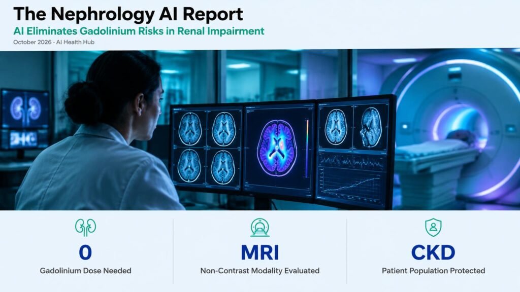
A multicohort diagnostic accuracy study published in The Lancet Medical Imaging & Theranostics demonstrates that deep learning models can predict brain tumor contrast enhancement using only non-contrast MRI sequences (The Lancet Medical Imaging & Theranostics). This advancement offers a potential alternative to gadolinium-based contrast agents for neuroimaging, directly addressing a critical clinical challenge in patients with severe renal impairment who face risks such as nephrogenic systemic fibrosis or systemic gadolinium retention.
Diagnostic Precision Without Chemical Contrast
Standard oncologic neuroimaging relies on post-contrast T1-weighted sequences to identify blood-brain barrier disruption and define tumor margins. However, for nephrologists managing patients with advanced chronic kidney disease (CKD) or acute kidney injury, authorizing contrast-enhanced MRI requires balancing diagnostic necessity against potential toxicities. The deep learning model synthesizes non-contrast MRI sequences to generate predicted contrast-enhanced images, mapping hyperintense tissue features that correspond to true pathological vascular permeability.
Clinical Limitations and Diagnostic Scope
While the multicohort retrospective design validates strong diagnostic accuracy across diverse patient cohorts, the study evaluated algorithmic image synthesis against traditional post-contrast scans rather than direct clinical patient outcomes. Retrospective validation on existing MRI datasets does not demonstrate that synthetic enhancement reliably guides surgical planning or radiation therapy in lieu of physical contrast agents. Additionally, non-contrast deep learning predictions depend on the underlying quality and artifact profile of baseline unenhanced scans, which vary across scanner manufacturers and field strengths.
Practical Takeaways for Nephrology Practice
This synthetic imaging approach represents a diagnostic accuracy milestone rather than an immediate replacement for clinical contrast protocols. Nephrologists and neuroradiologists should view non-contrast AI image synthesis as an emerging risk-mitigation tool for high-risk patients who require frequent follow-up neuroimaging but cannot safely clear gadolinium. Clinical adoption will require prospective interventional trials demonstrating that AI-generated enhancement produces equivalent therapeutic decisions without increasing diagnostic misclassification.